產(chǎn)品介紹 This gene encodes the enzyme responsible for RNA editing by site-specific deamination of adenosines. This enzyme destabilizes double-stranded RNA through conversion of adenosine to inosine. Mutations in this gene have been associated with dyschromatosis symmetrica hereditaria. Alternative splicing results in multiple transcript variants. [provided by RefSeq, Jul 2010]
Function:
Converts multiple adenosines to inosines and creates I/U mismatched base pairs in double-helical RNA substrates without apparent sequence specificity. Has been found to modify more frequently adenosines in AU-rich regions, probably due to the relative ease of melting A/U base pairs as compared to G/C pairs. Functions to modify viral RNA genomes and may be responsible for hypermutation of certain negative-stranded viruses. Edits the messenger RNAs for glutamate receptor (GLUR) subunits by site-selective adenosine deamination. Produces low-level editing at the GLUR-B Q/R site, but edits efficiently at the R/G site and HOTSPOT1. Binds to short interfering RNAs (siRNA) without editing them and suppresses siRNA-mediated RNA interference. Binds to ILF3/NF90 and up-regulates ILF3-mediated gene expression.
Subunit:
Homodimer. Isoform 1 interacts with ILF2/NF45 and ILF3/NF90.
Subcellular Location:
Cytoplasm. Nucleus, nucleolus. Isoform 1: Cytoplasm. Note=Found predominantly in cytoplasm but appears to shuttle between the cytoplasm and nucleus. Isoform 5: Nucleus, nucleolus.
Tissue Specificity:
Ubiquitously expressed, highest levels were found in brain and lung.
Post-translational modifications:
Sumoylation reduces RNA-editing activity.
DISEASE:
Defects in ADAR are a cause of dyschromatosis symmetrical hereditaria (DSH) [MIM:127400]; also known as reticulate acropigmentation of Dohi. DSH is a pigmentary genodermatosis of autosomal dominant inheritance characterized by a mixture of hyperpigmented and hypopigmented macules distributed on the dorsal parts of the hands and feet.
Similarity:
Contains 1 A to I editase domain.
Contains 2 DRADA
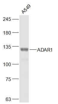
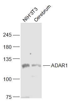
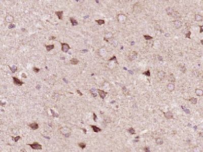
| 產(chǎn)品圖片 | 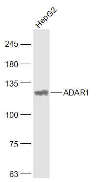 Sample: HepG2(Human) Cell Lysate at 30 ug Primary: Anti-ADAR1 (bs-2168R) at 1/1000 dilution Secondary: IRDye800CW Goat Anti-Rabbit IgG at 1/20000 dilution Predicted band size: 135 kD Observed band size: 110 kD Sample: A549(Human) Cell Lysate at 30 ug Primary: Anti-ADAR1 (bs-2168R) at 1/1000 dilution Secondary: IRDye800CW Goat Anti-Rabbit IgG at 1/20000 dilution Predicted band size: 135 kD Observed band size: 110 kD Sample: NIH/3T3(Mouse) Cell Lysate at 30 ug Cerebrum (Mouse) Lysate at 40 ug Primary: Anti-ADAR1 (bs-2168R) at 1/1000 dilution Secondary: IRDye800CW Goat Anti-Rabbit IgG at 1/20000 dilution Predicted band size: 135 kD Observed band size: 110 kD Paraformaldehyde-fixed, paraffin embedded (Rat brain); Antigen retrieval by boiling in sodium citrate buffer (pH6.0) for 15min; Block endogenous peroxidase by 3% hydrogen peroxide for 20 minutes; Blocking buffer (normal goat serum) at 37°C for 30min; Antibody incubation with (ADAR1) Polyclonal Antibody, Unconjugated (bs-2168R) at 1:400 overnight at 4°C, followed by a conjugated secondary antibody (sp-0023) for 20 minutes and DAB staining. |
我要詢價
*聯(lián)系方式:
(可以是QQ、MSN、電子郵箱、電話等,您的聯(lián)系方式不會被公開)
*內(nèi)容:


