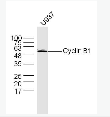
| 中文名稱 | 周期素B1抗體 |
| 別 名 | CCNB 1; CCNB; CCNB1; CCNB1_HUMAN; G2 mitotic specific cyclin B1; G2/mitotic-specific cyclin-B1. |
| 研究領域 | 腫瘤 細胞生物 免疫學 細胞周期蛋白 |
| 抗體來源 | Rabbit |
| 克隆類型 | Polyclonal |
| 交叉反應 | Human, Mouse, Rat, (predicted: Cow, ) |
| 產品應用 | WB=1:500-2000 ELISA=1:500-1000 IHC-P=1:100-500 IHC-F=1:100-500 Flow-Cyt=1μg/Test IF=1:100-500 (石蠟切片需做抗原修復) not yet tested in other applications. optimal dilutions/concentrations should be determined by the end user. |
| 分 子 量 | 48kDa |
| 細胞定位 | 細胞核 細胞漿 |
| 性 狀 | Liquid |
| 濃 度 | 1mg/ml |
| 免 疫 原 | KLH conjugated synthetic peptide derived from human Cyclin B1:271-433/433 |
| 亞 型 | IgG |
| 純化方法 | affinity purified by Protein A |
| 儲 存 液 | 0.01M TBS(pH7.4) with 1% BSA, 0.03% Proclin300 and 50% Glycerol. |
| 保存條件 | Shipped at 4℃. Store at -20 °C for one year. Avoid repeated freeze/thaw cycles. |
| PubMed | PubMed |
| 產品介紹 | The protein encoded by this gene is a regulatory protein involved in mitosis. The gene product complexes with p34(cdc2) to form the maturation-promoting factor (MPF). Two alternative transcripts have been found, a constitutively expressed transcript and a cell cycle-regulated transcript, that is expressed predominantly during G2/M phase. The different transcripts result from the use of alternate transcription initiation sites. [provided by RefSeq, Jul 2008]. Function: Essential for the control of the cell cycle at the G2/M (mitosis) transition. Subunit: Interacts with the CDC2 protein kinase to form a serine/threonine kinase holoenzyme complex also known as maturation promoting factor (MPF). The cyclin subunit imparts substrate specificity to the complex. Binds HEI10. Interacts with catalytically active RALBP1 and CDC2 during mitosis to form an endocytotic complex during interphase. Interacts with CCNF; interaction is required for nuclear localization. Subcellular Location: Cytoplasm. Nucleus. Cytoplasm, cytoskeleton, centrosome. Post-translational modifications: Ubiquitinated by the SCF(NIPA) complex during interphase, leading to its destruction. Not ubiquitinated during G2/M phases. Phosphorylated by PLK1 at Ser-133 on centrosomes during prophase: phosphorylation by PLK1 does not cause nuclear import. Phosphorylation at Ser-147 was also reported to be mediated by PLK1 but Ser-133 seems to be the primary phosphorylation site. Similarity: Belongs to the cyclin family. Cyclin AB subfamily. SWISS: P24860 Gene ID: 891 Database links: Entrez Gene: 891 Human Entrez Gene: 268697 Mouse Entrez Gene: 25203 Rat Omim: 123836 Human SwissProt: P14635 Human SwissProt: P24860 Mouse SwissProt: P30277 Rat Unigene: 23960 Human Unigene: 260114 Mouse Unigene: 380027 Mouse Unigene: 482545 Mouse Unigene: 9232 Rat Important Note: This product as supplied is intended for research use only, not for use in human, therapeutic or diagnostic applications. 主要出現在G2期。Cyclin B是細胞周期調節必不可少的條件。 細胞周期素B1是細胞周期調節因子,它的異常表達將導致細胞周期發生紊亂,致使腫瘤形成. |
| 產品圖片 | 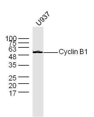 Sample: Sample:U937 Cell (Human) Lysate at 30 ug Primary: Anti- Cyclin B1 (bs-0572R) at 1/300 dilution Secondary: IRDye800CW Goat Anti-Rabbit IgG at 1/20000 dilution Predicted band size: 48 kD Observed band size: 50 kD 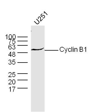 Sample: Sample:U251 Cell (Human) Lysate at 30 ug Primary: Anti- Cyclin B1 (bs-0572R) at 1/300 dilution Secondary: IRDye800CW Goat Anti-Rabbit IgG at 1/20000 dilution Predicted band size: 48 kD Observed band size: 50 kD 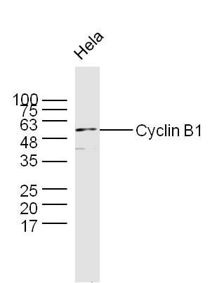 Sample: Sample:Hela Cell (Human) Lysate at 30 ug Primary: Anti- Cyclin B1 (bs-0572R) at 1/300 dilution Secondary: IRDye800CW Goat Anti-Rabbit IgG at 1/20000 dilution Predicted band size: 48 kD Observed band size: 50 kD 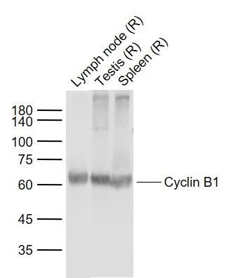 Sample: Sample:Lane 1: Lymph node (Rat) Lysate at 40 ug Lane 2: Testis (Rat) Lysate at 40 ug Lane 3: Spleen (Rat) Lysate at 40 ug Primary: Anti-Cyclin B1 (bs-0572R) at 1/1000 dilution Secondary: IRDye800CW Goat Anti-Rabbit IgG at 1/20000 dilution Predicted band size: 55-60 kD Observed band size: 60 kD 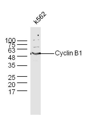 Sample: Sample:K562 Cell (Human) Lysate at 30 ug Primary: Anti- Cyclin B1 (bs-0572R) at 1/300 dilution Secondary: IRDye800CW Goat Anti-Rabbit IgG at 1/20000 dilution Predicted band size: 48 kD Observed band size: 50 kD 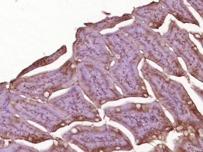 Paraformaldehyde-fixed, paraffin embedded (Mouse small intestine); Antigen retrieval by boiling in sodium citrate buffer (pH6.0) for 15min; Block endogenous peroxidase by 3% hydrogen peroxide for 20 minutes; Blocking buffer (normal goat serum) at 37°C for 30min; Antibody incubation with (Cyclin B1) Polyclonal Antibody, Unconjugated (bs-0572R) at 1:400 overnight at 4°C, followed by operating according to SP Kit(Rabbit) (sp-0023) instructionsand DAB staining. Paraformaldehyde-fixed, paraffin embedded (Mouse small intestine); Antigen retrieval by boiling in sodium citrate buffer (pH6.0) for 15min; Block endogenous peroxidase by 3% hydrogen peroxide for 20 minutes; Blocking buffer (normal goat serum) at 37°C for 30min; Antibody incubation with (Cyclin B1) Polyclonal Antibody, Unconjugated (bs-0572R) at 1:400 overnight at 4°C, followed by operating according to SP Kit(Rabbit) (sp-0023) instructionsand DAB staining.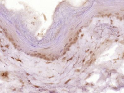 Paraformaldehyde-fixed, paraffin embedded (Rat esophageal); Antigen retrieval by boiling in sodium citrate buffer (pH6.0) for 15min; Block endogenous peroxidase by 3% hydrogen peroxide for 20 minutes; Blocking buffer (normal goat serum) at 37°C for 30min; Antibody incubation with (Cyclin B1) Polyclonal Antibody, Unconjugated (bs-0572R) at 1:400 overnight at 4°C, followed by operating according to SP Kit(Rabbit) (sp-0023) instructionsand DAB staining. Paraformaldehyde-fixed, paraffin embedded (Rat esophageal); Antigen retrieval by boiling in sodium citrate buffer (pH6.0) for 15min; Block endogenous peroxidase by 3% hydrogen peroxide for 20 minutes; Blocking buffer (normal goat serum) at 37°C for 30min; Antibody incubation with (Cyclin B1) Polyclonal Antibody, Unconjugated (bs-0572R) at 1:400 overnight at 4°C, followed by operating according to SP Kit(Rabbit) (sp-0023) instructionsand DAB staining.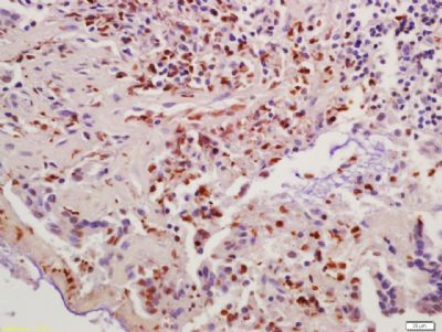 Tissue/cell: human colon carcinoma; 4% Paraformaldehyde-fixed and paraffin-embedded; Tissue/cell: human colon carcinoma; 4% Paraformaldehyde-fixed and paraffin-embedded;Antigen retrieval: citrate buffer ( 0.01M, pH 6.0 ), Boiling bathing for 15min; Block endogenous peroxidase by 3% Hydrogen peroxide for 30min; Blocking buffer (normal goat serum,C-0005) at 37℃ for 20 min; Incubation: Anti-Cyclin B1 Polyclonal Antibody, Unconjugated(bs-0572R) 1:200, overnight at 4°C, followed by conjugation to the secondary antibody(SP-0023) and DAB(C-0010) staining 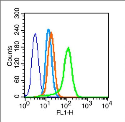 Blank control (blue line): A549 (blue). Blank control (blue line): A549 (blue).Primary Antibody (green line): Rabbit Anti-Cyclin B1 antibody(bs-0572R). Dilution: 1μg /10^6 cells; Isotype Control Antibody (orange line): Rabbit IgG . Secondary Antibody (white blue line): F(ab’)2 fragment goat anti-rabbit IgG-FITC. Dilution: 1μg /test. Protocol The cells were fixed with 2% paraformaldehyde (10 min) and then permeabilized with 0.1% PBS-Tween for 20 min at room temperature.Cells stained with Primary Antibody for 30 min at room temperature. The cells were then incubated in 1 X PBS/2%BSA/10% goat serum to block non-specific protein-protein interactions followed by the antibody for 15 min at room temperature. The secondary antibody used for 40 min at room temperature. Acquisition of 20,000 events was performed. 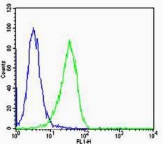 Cell: Hela Cell: HelaConcentration:1:100 Host/Isotype:Rabbit/IgG Flow cytometric analysis of primary antibody (Cat#: bs-0572R) on Hela(green) compared with isotype control in the absence of primary antibody (blue) followed by Alexa Fluor 488-conjugated goat anti-rabbit IgG(H+L) secondary antibody . |
我要詢價
*聯系方式:
(可以是QQ、MSN、電子郵箱、電話等,您的聯系方式不會被公開)
*內容:


