| 中文名稱 | FasL抗體 |
| 別 名 | APO1L; Apoptosis (APO 1) antigen ligand 1; Fas-L; Apoptosis antigen ligand 1; APTL; CD178; CD-178; CD 178; CD95L; CD95 ligand; CD95-L; CD95L protein; FAS antigen ligand; Fas L; Fas Ligand; FASL; Fasl; Fas ligand (TNF superfamily member 6); Generalized lymphoproliferative disease; TNF superfamily member 6; TNFSF6; Tumor necrosis factor ligand superfamily member 6; TNFL6_HUMAN; Apoptosis antigen ligand; Tumor necrosis factor ligand superfamily member 6, membrane form; Tumor necrosis factor ligand superfamily member 6, soluble form; FASLG. |
| 研究領域 | 腫瘤 細胞生物 免疫學 細胞凋亡 |
| 抗體來源 | Rabbit |
| 克隆類型 | Polyclonal |
| 交叉反應 | Human, Mouse, Rat, (predicted: Cow, ) |
| 產品應用 | WB=1:500-2000 ELISA=1:500-1000 IHC-P=1:100-500 IHC-F=1:100-500 Flow-Cyt=1μg/Test ICC=1:100-500 IF=1:100-500 (石蠟切片需做抗原修復) not yet tested in other applications. optimal dilutions/concentrations should be determined by the end user. |
| 分 子 量 | 31kDa |
| 細胞定位 | 細胞核 細胞膜 細胞外基質 分泌型蛋白 |
| 性 狀 | Liquid |
| 濃 度 | 1mg/ml |
| 免 疫 原 | KLH conjugated synthetic peptide derived from human Fas Ligand:196-281/281 |
| 亞 型 | IgG |
| 純化方法 | affinity purified by Protein A |
| 儲 存 液 | 0.01M TBS(pH7.4) with 1% BSA, 0.03% Proclin300 and 50% Glycerol. |
| 保存條件 | Shipped at 4℃. Store at -20 °C for one year. Avoid repeated freeze/thaw cycles. |
| PubMed | PubMed |
| 產品介紹 | FasL (CD95L) is a cytokine that binds to TNFRSF6/FAS, a receptor that transduces the apoptotic signal into cells. May be involved in cytotoxic T cell mediated apoptosis and in T cell development. TNFRSF6/FAS-mediated apoptosis may have a role in the induction of peripheral tolerance, in the antigen-stimulated suicide of mature T cells, or both. Binding to the decoy receptor TNFRSF6B/DcR3 modulates its effects. Homotrimer (Probable). May be released as type II membrane protein. Belongs to the tumor necrosis factor family Function: Cytokine that binds to TNFRSF6/FAS, a receptor that transduces the apoptotic signal into cells. May be involved in cytotoxic T-cell mediated apoptosis and in T-cell development. TNFRSF6/FAS-mediated apoptosis may have a role in the induction of peripheral tolerance, in the antigen-stimulated suicide of mature T-cells, or both. Binding to the decoy receptor TNFRSF6B/DcR3 modulates its effects. Subunit: Homotrimer (Probable). Interacts with ARHGAP9, BAIAP2L1, BTK, CACNB3, CACNB4, CRK, DLG2, DNMBP, DOCK4, EPS8L3, FGR, FYB, FYN, HCK, ITK, ITSN2, KALRN, LYN, MACC1, MIA, MPP4, MYO15A, NCF1, NCK1, NCK2, NCKIPSD, OSTF1, PIK3R1, PSTPIP1, RIMBP3C, SAMSN1, SH3GL3, SH3PXD2B, SH3PXD2A, SH3RF2, SKAP2, SNX33, SNX9, SORBS3, SPTA1, SRC, SRGAP1, SRGAP2, SRGAP3, TEC, TJP3 and YES1. Subcellular Location: Cell membrane; Single-pass type II membrane protein. Secreted. Cytoplasmic vesicle lumen. Lysosome lumen. Note=May be released into the extracellular fluid, probably by cleavage form the cell surface. Is internalized into multivesicular bodies of secretory lysosomes after phosphorylation by FGR and monoubiquitination. Post-translational modifications: N-glycosylated. The soluble form derives from the membrane form by proteolytic processing. Phosphorylated by FGR on tyrosine residues; this is required for ubiquitination and subsequent internalization. Monoubiquitinated. DISEASE: Defects in FASLG are the cause of autoimmune lymphoproliferative syndrome type 1B (ALPS1B) [MIM:601859]; also known as Canale-Smith syndrome (CSS). ALPS is a childhood syndrome involving hemolytic anemia and thrombocytopenia with massive lymphadenopathy and splenomegaly. Similarity: Belongs to the tumor necrosis factor family. SWISS: P48023 Gene ID: 356 Database links: Entrez Gene: 356 Human Entrez Gene: 14103 Mouse Entrez Gene: 25385 Rat Omim: 134638 Human SwissProt: P48023 Human SwissProt: P41047 Mouse SwissProt: P36940 Rat Unigene: 2007 Human Unigene: 3355 Mouse Unigene: 9725 Rat Important Note: This product as supplied is intended for research use only, not for use in human, therapeutic or diagnostic applications. Fas-L 是一個與TNFRSF6/FAS 結合的細胞因子,一個轉導凋亡信號到細胞內的一個受體。他可能參與T-細胞調節凋亡細胞毒性和T-細胞的發育。調節凋亡的TNFRSF6/FAS 在外周組織耐受性方面和抗原刺激成熟淋巴細胞自殺中起作用。FasL誘導物受體TNFRSF6B/DcR3結合調節其作用。 FasL只表達于活化的淋巴細胞,但在小鼠睪丸中可見高表達的FasL;分子量為36-43kDa。屬于TNF家族。 |
| 產品圖片 | 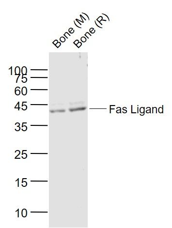 Sample: Sample:Lane 1: Bone (Mouse) Lysate at 40 ug Lane 2: Bone (Rat) Lysate at 40 ug Primary: Anti-Fas Ligand (bs-0216R) at 1/1000 dilution Secondary: IRDye800CW Goat Anti-Rabbit IgG at 1/20000 dilution Predicted band size: 40 kD Observed band size: 40 kD 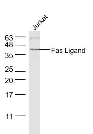 Sample: Sample:Jurkat(Human) Cell Lysate at 30 ug Primary: Anti-Fas Ligand (bs-0216R) at 1/1000 dilution Secondary: IRDye800CW Goat Anti-Rabbit IgG at 1/20000 dilution Predicted band size: 31 kD Observed band size: 31 kD 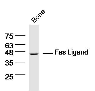 Sample: Bone (mouse) Lysate at 40 ug Sample: Bone (mouse) Lysate at 40 ugPrimary: Anti- Fas ligand (bs-0216R) at 1/300 dilution Secondary: IRDye800CW Goat Anti-Rabbit IgG at 1/20000 dilution Predicted band size: 31 kD Observed band size: 46 kD 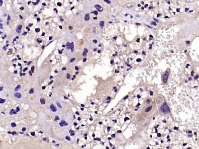 Paraformaldehyde-fixed, paraffin embedded (mouse placenta); Antigen retrieval by boiling in sodium citrate buffer (pH6.0) for 15min; Block endogenous peroxidase by 3% hydrogen peroxide for 20 minutes; Blocking buffer (normal goat serum) at 37°C for 30min; Antibody incubation with (Fas Ligand) Polyclonal Antibody, Unconjugated (bs-0216R) at 1:200 overnight at 4°C, followed by operating according to SP Kit(Rabbit) (sp-0023) instructionsand DAB staining. Paraformaldehyde-fixed, paraffin embedded (mouse placenta); Antigen retrieval by boiling in sodium citrate buffer (pH6.0) for 15min; Block endogenous peroxidase by 3% hydrogen peroxide for 20 minutes; Blocking buffer (normal goat serum) at 37°C for 30min; Antibody incubation with (Fas Ligand) Polyclonal Antibody, Unconjugated (bs-0216R) at 1:200 overnight at 4°C, followed by operating according to SP Kit(Rabbit) (sp-0023) instructionsand DAB staining.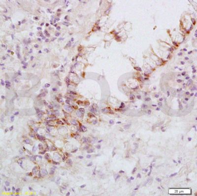 Tissue/cell: human rectal carcinoma; 4% Paraformaldehyde-fixed and paraffin-embedded; Tissue/cell: human rectal carcinoma; 4% Paraformaldehyde-fixed and paraffin-embedded;Antigen retrieval: citrate buffer ( 0.01M, pH 6.0 ), Boiling bathing for 15min; Block endogenous peroxidase by 3% Hydrogen peroxide for 30min; Blocking buffer (normal goat serum,C-0005) at 37℃ for 20 min; Incubation: Anti-FasL Polyclonal Antibody, Unconjugated(bs-0216R) 1:200, overnight at 4°C, followed by conjugation to the secondary antibody(SP-0023) and DAB(C-0010) staining 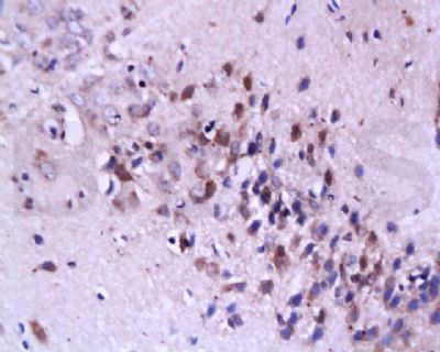 Tissue/cell: rat brain tissue; 4% Paraformaldehyde-fixed and paraffin-embedded; Tissue/cell: rat brain tissue; 4% Paraformaldehyde-fixed and paraffin-embedded;Antigen retrieval: citrate buffer ( 0.01M, pH 6.0 ), Boiling bathing for 15min; Block endogenous peroxidase by 3% Hydrogen peroxide for 30min; Blocking buffer (normal goat serum,C-0005) at 37℃ for 20 min; Incubation: Anti-FasL Polyclonal Antibody, Unconjugated(bs-0216R) 1:200, overnight at 4°C, followed by conjugation to the secondary antibody(SP-0023) and DAB(C-0010) staining 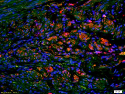 Tissue/cell: human rectal carcinoma;4% Paraformaldehyde-fixed and paraffin-embedded; Tissue/cell: human rectal carcinoma;4% Paraformaldehyde-fixed and paraffin-embedded;Antigen retrieval: citrate buffer ( 0.01M, pH 6.0 ), Boiling bathing for 15min; Blocking buffer (normal goat serum,C-0005) at 37℃ for 20 min; Incubation: Anti-FasL Polyclonal Antibody, Unconjugated(bs-0216R) 1:200, overnight at 4°C; The secondary antibody was Goat Anti-Rabbit IgG, Cy3 conjugated(bs-0295G-Cy3)used at 1:200 dilution for 40 minutes at 37°C. DAPI(5ug/ml,blue,C-0033) was used to stain the cell nuclei  Blank control: Mouse Kidney (blue). Blank control: Mouse Kidney (blue).Primary Antibody:Rabbit Anti-phospho-Fas Ligand antibody (bs-0216R,Green); Dilution: 1μg in 100 μL 1X PBS containing 0.5% BSA; Isotype Control Antibody: Rabbit IgG(orange) ,used under the same conditions; Secondary Antibody: Goat anti-rabbit IgG-FITC(white blue), Dilution: 1:200 in 1 X PBS containing 0.5% BSA. Protocol The cells were fixed with 2% paraformaldehyde for 10 min at 37℃. Primary antibody (bs-0216R, 1μg /1x10^6 cells) were incubated for 30 min at room temperature, followed by 1 X PBS containing 0.5% BSA + 1 0% goat serum (15 min) to block non-specific protein-protein interactions. Then the Goat Anti-rabbit IgG/FITC antibody was added into the blocking buffer mentioned above to react with the primary antibody at 1/200 dilution for 40 min on ice. Acquisition of 20,000 events was performed. 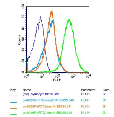 Blank control: mouse thymouses(blue) Blank control: mouse thymouses(blue)Isotype Control Antibody: Rabbit IgG(orange) ; Secondary Antibody: Goat anti-rabbit IgG-FITC(white blue), Dilution: 1:100 in 1 X PBS containing 0.5% BSA ; Primary Antibody Dilution: 1μl in 100 μL1X PBS containing 0.5% BSA(green). |
我要詢價
*聯系方式:
(可以是QQ、MSN、電子郵箱、電話等,您的聯系方式不會被公開)
*內容:


