| 英文名稱 | MCL1 |
| 中文名稱 | 髓樣細胞白血病-1抗體 |
| 別 名 | myeloid cell leukemia 1; myeloid cell leukemia sequence 1; MCL-1; MCL1L; MCL 1; mcl1/EAT; MGC104264; MGC1839; TM; MCL1S; EAT) Bcl 2 related protein EAT/mcl1; BCL2 related; BCL2L3; EAT; Induced myeloid leukemia cell differentiation protein Mcl 1; myeloid cell leukemia sequence 1; Myeloid cell leukemia sequence 1 BCL2 related; Myeloid cell leukemia sequence 1 isoform 1; OTTHUMP00000032794; OTTHUMP00000032795; TM; bcl2-L-3; BCL2L3; EAT; Mcl-1; MCL1-ES; mcl1/EAT. |
| 研究領域 | 腫瘤 免疫學 信號轉導 細胞凋亡 轉錄調節因子 線粒體 |
| 抗體來源 | Rabbit |
| 克隆類型 | Polyclonal |
| 交叉反應 | Human, Mouse, Rat, (predicted: Dog, Pig, Horse, Rabbit, ) |
| 產品應用 | WB=1:500-2000 IHC-P=1:100-500 IHC-F=1:100-500 ICC=1:100-500 IF=1:100-500 (石蠟切片需做抗原修復) not yet tested in other applications. optimal dilutions/concentrations should be determined by the end user. |
| 分 子 量 | 39kDa |
| 細胞定位 | 細胞核 細胞漿 細胞膜 線粒體 |
| 性 狀 | Liquid |
| 濃 度 | 1mg/ml |
| 免 疫 原 | KLH conjugated synthetic peptide derived from human MCL1:101-200/350 |
| 亞 型 | IgG |
| 純化方法 | affinity purified by Protein A |
| 儲 存 液 | 0.01M TBS(pH7.4) with 1% BSA, 0.03% Proclin300 and 50% Glycerol. |
| 保存條件 | Shipped at 4℃. Store at -20 °C for one year. Avoid repeated freeze/thaw cycles. |
| PubMed | PubMed |
| 產品介紹 | Mcl1 is an anti-apoptotic member of Bcl2 family originally isolated from the ML1 human myeloid leukemia cell line during phorbol ester-induced differentiation along the monocyte/macrophage pathway. Mcl1 localizes to the mitochondria, interacts with and antagonizes pro-apoptotic Bcl2 family members, and inhibits apoptosis by a number of cytotoxic stimuli. It is involved in programing of differentiation and concomitant maintenance of viability but not of proliferation. Isoform 1 inhibits apoptosis while isoform 2 promotes it. Expression increases early during phorbol-ester induced differentiation along the monocyte/macrophage pathway in myeloid leukemia cell lines ML1. Function: Involved in the regulation of apoptosis versus cell survival, and in the maintenance of viability but not of proliferation. Mediates its effects by interactions with a number of other regulators of apoptosis. Isoform 1 inhibits apoptosis. Isoform 2 promotes apoptosis. Subunit: Interacts with BAD, BOK, BIK and BFM (By similarity). Interacts with PMAIP1. Isoform 1 interacts with BAX, BAK1, TPT1 and BCL2L11. Heterodimer of isoform 1 and isoform 2. Homodimers of isoform 1 or isoform 2 are not detected. Isoform 2 does not interact with pro-apoptotic BCL2-related proteins. Subcellular Location: Membrane; Single-pass membrane protein (Potential). Cytoplasm. Mitochondrion. Nucleus, nucleoplasm. Note=Cytoplasmic, associated with mitochondria. Post-translational modifications: Cleaved by CASP3 during apoptosis. In intact cells cleavage occurs preferentially after Asp-127, yielding a pro-apoptotic 28 kDa C-terminal fragment. Rapidly degraded in the absence of phosphorylation on Thr-163 in the PEST region. Phosphorylated on Thr-163. Treatment with taxol or okadaic acid induces phosphorylation on additional sites. Similarity: Belongs to the Bcl-2 family. SWISS: Q07820 Gene ID: 4170 Database links: Entrez Gene: 4170 Human Entrez Gene: 17210 Mouse Entrez Gene: 60430 Rat Omim: 159552 Human SwissProt: Q07820 Human SwissProt: P97287 Mouse SwissProt: Q9Z1P3 Rat Unigene: 632486 Human Important Note: This product as supplied is intended for research use only, not for use in human, therapeutic or diagnostic applications. |
| 產品圖片 | 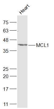 Sample: Sample:Heart (Rat) Lysate at 40 ug Primary: Anti-MCL1 (bs-23315R) at 1/1000 dilution Secondary: IRDye800CW Goat Anti-Rabbit IgG at 1/20000 dilution Predicted band size: 39 kD Observed band size: 41 kD 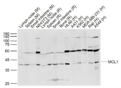 Sample: Sample:Lane 1: Lymph node (Mouse) Lysate at 40 ug Lane 2: Spleen (Mouse) Lysate at 40 ug Lane 3: Small intestine (Mouse) Lysate at 40 ug Lane 4: NIH/3T3 (Mouse) Cell Lysate at 30 ug Lane 5: Lymph node (Rat) Lysate at 40 ug Lane 6: Spleen (Rat) Lysate at 40 ug Lane 7: Small intestine (Rat) Lysate at 40 ug Lane 8: Bone (Rat) Lysate at 40 ug Lane 9: HL60 (Human) Cell Lysate at 30 ug Lane 10: A431 (Human) Cell Lysate at 30 ug Lane 11: K562 (Human) Cell Lysate at 30 ug Lane 12: MDA-MB-231 (Human) Cell Lysate at 30 ug Lane 13: Raji (Human) Cell Lysate at 30 ug Lane 14: A549 (Human) Cell Lysate at 30 ug Primary: Anti-MCL1 (bs-23315R) at 1/1000 dilution Secondary: IRDye800CW Goat Anti-Rabbit IgG at 1/20000 dilution Predicted band size: 39 kD Observed band size: 40 kD 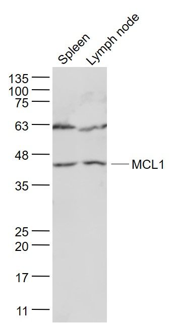 Sample: Sample:Spleen (Mouse) Lysate at 40 ug Lymph node (Mouse) Lysate at 40 ug Primary: Anti- MCL1 (bs-23315R) at 1/1000 dilution Secondary: IRDye800CW Goat Anti-Rabbit IgG at 1/20000 dilution Predicted band size: 39 kD Observed band size: 39 kD 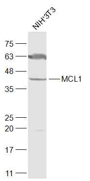 Sample: Sample:NIH/3T3(Mouse) Cell Lysate at 30 ug Primary: Anti-MCL1 (bs-23315R) at 1/1000 dilution Secondary: IRDye800CW Goat Anti-Rabbit IgG at 1/20000 dilution Predicted band size: 39 kD Observed band size: 41 kD 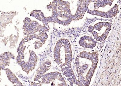 Paraformaldehyde-fixed, paraffin embedded (human colon carcinoma); Antigen retrieval by boiling in sodium citrate buffer (pH6.0) for 15min; Block endogenous peroxidase by 3% hydrogen peroxide for 20 minutes; Blocking buffer (normal goat serum) at 37°C for 30min; Antibody incubation with (MCL1) Polyclonal Antibody, Unconjugated (bs-23315R) at 1:200 overnight at 4°C, followed by operating according to SP Kit(Rabbit) (sp-0023) instructionsand DAB staining. Paraformaldehyde-fixed, paraffin embedded (human colon carcinoma); Antigen retrieval by boiling in sodium citrate buffer (pH6.0) for 15min; Block endogenous peroxidase by 3% hydrogen peroxide for 20 minutes; Blocking buffer (normal goat serum) at 37°C for 30min; Antibody incubation with (MCL1) Polyclonal Antibody, Unconjugated (bs-23315R) at 1:200 overnight at 4°C, followed by operating according to SP Kit(Rabbit) (sp-0023) instructionsand DAB staining.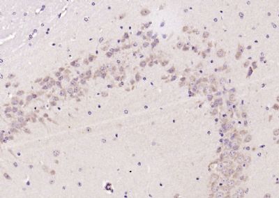 Paraformaldehyde-fixed, paraffin embedded (mouse brain); Antigen retrieval by boiling in sodium citrate buffer (pH6.0) for 15min; Block endogenous peroxidase by 3% hydrogen peroxide for 20 minutes; Blocking buffer (normal goat serum) at 37°C for 30min; Antibody incubation with (MCL1) Polyclonal Antibody, Unconjugated (bs-23315R) at 1:200 overnight at 4°C, followed by operating according to SP Kit(Rabbit) (sp-0023) instructionsand DAB staining. Paraformaldehyde-fixed, paraffin embedded (mouse brain); Antigen retrieval by boiling in sodium citrate buffer (pH6.0) for 15min; Block endogenous peroxidase by 3% hydrogen peroxide for 20 minutes; Blocking buffer (normal goat serum) at 37°C for 30min; Antibody incubation with (MCL1) Polyclonal Antibody, Unconjugated (bs-23315R) at 1:200 overnight at 4°C, followed by operating according to SP Kit(Rabbit) (sp-0023) instructionsand DAB staining.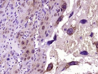 Paraformaldehyde-fixed, paraffin embedded (Mouse embryo); Antigen retrieval by boiling in sodium citrate buffer (pH6.0) for 15min; Block endogenous peroxidase by 3% hydrogen peroxide for 20 minutes; Blocking buffer (normal goat serum) at 37°C for 30min; Antibody incubation with (MCL1) Polyclonal Antibody, Unconjugated (bs-23315R) at 1:400 overnight at 4°C, followed by operating according to SP Kit(Rabbit) (sp-0023) instructions and DAB staining. Paraformaldehyde-fixed, paraffin embedded (Mouse embryo); Antigen retrieval by boiling in sodium citrate buffer (pH6.0) for 15min; Block endogenous peroxidase by 3% hydrogen peroxide for 20 minutes; Blocking buffer (normal goat serum) at 37°C for 30min; Antibody incubation with (MCL1) Polyclonal Antibody, Unconjugated (bs-23315R) at 1:400 overnight at 4°C, followed by operating according to SP Kit(Rabbit) (sp-0023) instructions and DAB staining.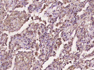 Paraformaldehyde-fixed, paraffin embedded (human lung carcinoma); Antigen retrieval by boiling in sodium citrate buffer (pH6.0) for 15min; Block endogenous peroxidase by 3% hydrogen peroxide for 20 minutes; Blocking buffer (normal goat serum) at 37°C for 30min; Antibody incubation with (MCL1) Polyclonal Antibody, Unconjugated (bs-23315R) at 1:400 overnight at 4°C, followed by operating according to SP Kit(Rabbit) (sp-0023) instructionsand DAB staining. Paraformaldehyde-fixed, paraffin embedded (human lung carcinoma); Antigen retrieval by boiling in sodium citrate buffer (pH6.0) for 15min; Block endogenous peroxidase by 3% hydrogen peroxide for 20 minutes; Blocking buffer (normal goat serum) at 37°C for 30min; Antibody incubation with (MCL1) Polyclonal Antibody, Unconjugated (bs-23315R) at 1:400 overnight at 4°C, followed by operating according to SP Kit(Rabbit) (sp-0023) instructionsand DAB staining. |
我要詢價
*聯系方式:
(可以是QQ、MSN、電子郵箱、電話等,您的聯系方式不會被公開)
*內容:


