| 中文名稱 | 原癌基因K-ras抗體 |
| 別 名 | C-K-RAS; c-Ki-ras; c-Ki-ras p21; Ha-ras; K-RAS B; K-RAS2A; K-RAS2B; K-RAS4A; KI-RAS; KI-RAS4B; KRAS; KRAS1; KRAS2; MGC7141; NS; NS3; p21; p21B; p21ras; RAS; RAS1; RASH; RASK2. |
| 研究領域 | 腫瘤 細胞生物 免疫學 信號轉導 細胞凋亡 細胞膜受體 轉運蛋白 |
| 抗體來源 | Rabbit |
| 克隆類型 | Polyclonal |
| 交叉反應 | Human, Mouse, Rat, |
| 產品應用 | WB=1:500-2000 ELISA=1:500-1000 IHC-P=1:100-500 IHC-F=1:100-500 Flow-Cyt=1ug/Tset IF=1:100-500 (石蠟切片需做抗原修復) not yet tested in other applications. optimal dilutions/concentrations should be determined by the end user. |
| 分 子 量 | 21kDa |
| 細胞定位 | 細胞核 細胞漿 細胞膜 |
| 性 狀 | Liquid |
| 濃 度 | 1mg/ml |
| 免 疫 原 | KLH conjugated synthetic peptide derived from human K-ras:25-130/189 |
| 亞 型 | IgG |
| 純化方法 | affinity purified by Protein A |
| 儲 存 液 | 0.01M TBS(pH7.4) with 1% BSA, 0.03% Proclin300 and 50% Glycerol. |
| 保存條件 | Shipped at 4℃. Store at -20 °C for one year. Avoid repeated freeze/thaw cycles. |
| PubMed | PubMed |
| 產品介紹 | This gene, a Kirsten ras oncogene homolog from the mammalian ras gene family, encodes a protein that is a member of the small GTPase superfamily. A single amino acid substitution is responsible for an activating mutation. The transforming protein that results is implicated in various malignancies, including lung adenocarcinoma, mucinous adenoma, ductal carcinoma of the pancreas and colorectal carcinoma. Alternative splicing leads to variants encoding two isoforms that differ in the C-terminal region. [provided by RefSeq] Ras, a proto-oncogene, is a small G-protein that has 3 primary isoforms (H-Ras, N-Ras, and K-Ras) that differ in there approximately 20 C-terminal amino acids. H-Ras was first discovered as a transforming product the retrovirus Harvey murine virus and K-Ras of Kirten sarcoma virus. Ras is a heavily studied target of both academic and pharmaceutical research because of its implications in various pathways and diseases as well as being mutated in a large number of human cancers. Ras is most notably the activator of the Erk/MAPK kinase pathway as activator of Raf, as well as an activator of PI3 Kinase (PI3K). In its oncogenic, mutated state, Ras is unable to hydrolyze GTP to GDP, thus staying in an active state and activating numerous pathways including the MAPK pathway through its activation of Raf, but also others as well that include PI3 Kinase and RalGDS. One path that the pharmaceutical industry has taken to control Ras and its activity is by finding what some consider its Achilles’ heel. For its activation, Ras must localize to the plasma membrane, but interestingly, it lacks a transmembrane domain. To achieve this, Ras must first undergo a post-translational modification (PTM) known as prenylation or geranylation at its C-terminal CAAX motif. For this to take place, a controlled three step process must occur. The first step in the process is the prenylation or geranylation of the C in the CAAX motif that is initiated by the covalent attachment of farnesyl groups to the cysteine that is catalyzed by the . After this modification, the and heterodimer enzymes farnesyl transferases –aaX of the motif is proteolytically removed via Rce1 (Ras Converting Enzyme 1), a membrane associated endoprotease, by a mechanism that is still not fully understood. Finally, the C-terminal prenylcysteine is now methlylated by ICMT (Isoprenylcysteine Carboxymethyl Transferase). These drugs have yet to pass clinical trials though and there is doubt that they will ever be successful in treating tumors associated with Ras activation. Function: Ras proteins bind GDP/GTP and possess intrinsic GTPase activity. Subunit: In its GTP-bound form interacts with PLCE1. Interacts with TBC1D10C. Interacts with RGL3. Interacts with HSPD1. Found in a complex with at least BRAF, HRAS1, MAP2K1, MAPK3 and RGS14. Interacts (active GTP-bound form) with RGS14 (via RBD 1 domain). Forms a signaling complex with RASGRP1 and DGKZ. Interacts with RASSF5. Interacts with PDE6D. Interacts with IKZF3. Interacts with GNB2L1. Subcellular Location: Cell membrane. Cell membrane; Lipid-anchor; Cytoplasmic side. Golgi apparatus. Golgi apparatus membrane; Lipid-anchor. Isoform 2: Nucleus. Cytoplasm. Cytoplasm, perinuclear region. Tissue Specificity: Widely expressed. DISEASE: Defects in KRAS are a cause of acute myelogenous leukemia (AML) [MIM:601626]. AML is a malignant disease in which hematopoietic precursors are arrested in an early stage of development. Defects in KRAS are a cause of juvenile myelomonocytic leukemia (JMML) [MIM:607785]. JMML is a pediatric myelodysplastic syndrome that constitutes approximately 30% of childhood cases of myelodysplastic syndrome (MDS) and 2% of leukemia. It is characterized by leukocytosis with tissue infiltration and in vitro hypersensitivity of myeloid progenitors to granulocyte-macrophage colony stimulating factor. Defects in KRAS are the cause of Noonan syndrome type 3 (NS3) [MIM:609942]. Noonan syndrome (NS) [MIM:163950] is a disorder characterized by dysmorphic facial features, short stature, hypertelorism, cardiac anomalies, deafness, motor delay, and a bleeding diathesis. It is a genetically heterogeneous and relatively common syndrome, with an estimated incidence of 1 in 1000-2500 live births. Rarely, NS is associated with juvenile myelomonocytic leukemia (JMML). NS3 inheritance is autosomal dominant. Defects in KRAS are a cause of gastric cancer (GASC) [MIM:613659]; also called gastric cancer intestinal or stomach cancer. Gastric cancer is a malignant disease which starts in the stomach, can spread to the esophagus or the small intestine, and can extend through the stomach wall to nearby lymph nodes and organs. It also can metastasize to other parts of the body. The term gastric cancer or gastric carcinoma refers to adenocarcinoma of the stomach that accounts for most of all gastric malignant tumors. Two main histologic types are recognized, diffuse type and intestinal type carcinomas. Diffuse tumors are poorly differentiated infiltrating lesions, resulting in thickening of the stomach. In contrast, intestinal tumors are usually exophytic, often ulcerating, and associated with intestinal metaplasia of the stomach, most often observed in sporadic disease. Note=Defects in KRAS are a cause of pylocytic astrocytoma (PA). Pylocytic astrocytomas are neoplasms of the brain and spinal cord derived from glial cells which vary from histologically benign forms to highly anaplastic and malignant tumors. Defects in KRAS are a cause of cardiofaciocutaneous syndrome (CFC syndrome) [MIM:115150]; also known as cardio-facio-cutaneous syndrome. CFC syndrome is characterized by a distinctive facial appearance, heart defects and mental retardation. Heart defects include pulmonic stenosis, atrial septal defects and hypertrophic cardiomyopathy. Some affected individuals present with ectodermal abnormalities such as sparse, friable hair, hyperkeratotic skin lesions and a generalized ichthyosis-like condition. Typical facial features are similar to Noonan syndrome. They include high forehead with bitemporal constriction, hypoplastic supraorbital ridges, downslanting palpebral fissures, a depressed nasal bridge, and posteriorly angulated ears with prominent helices. The inheritance of CFC syndrome is autosomal dominant. Note=KRAS mutations are involved in cancer development. Similarity: Belongs to the small GTPase superfamily. Ras family. SWISS: P01116 Gene ID: 3845 Database links: Entrez Gene: 3845 Human Entrez Gene: 16653 Mouse Entrez Gene: 24525 Rat Omim: 190070 Human SwissProt: P01116 Human SwissProt: P32883 Mouse SwissProt: P08644 Rat Unigene: 37003 Human Unigene: 505033 Human Unigene: 383182 Mouse Unigene: 24554 Rat Important Note: This product as supplied is intended for research use only, not for use in human, therapeutic or diagnostic applications. K-ras癌變基因的表達產物Ras蛋白存在于多數腫瘤之中,目前是腫瘤研究較重要的蛋白之一。 |
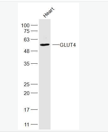
| 產品圖片 |  Sample: Sample:HL60(Human) Cell Lysate at 30 ug Primary: Anti- KRAS (bs-1033R) at 1/1000 dilution Secondary: IRDye800CW Goat Anti-Rabbit IgG at 1/20000 dilution Predicted band size: 21 kD Observed band size: 21 kD  Sample: Sample:NIH/3T3(Mouse) Cell Lysate at 30 ug Primary: Anti-KRAS (bs-1033R) at 1/300 dilution Secondary: IRDye800CW Goat Anti-Rabbit IgG at 1/20000 dilution Predicted band size: 21 kD Observed band size: 23 kD 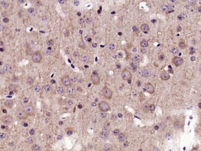 Paraformaldehyde-fixed, paraffin embedded (Mouse brain); Antigen retrieval by boiling in sodium citrate buffer (pH6.0) for 15min; Block endogenous peroxidase by 3% hydrogen peroxide for 20 minutes; Blocking buffer (normal goat serum) at 37°C for 30min; Antibody incubation with (KRAS) Polyclonal Antibody, Unconjugated (bs-1033R) at 1:400 overnight at 4°C, followed by operating according to SP Kit(Rabbit) (sp-0023) instructionsand DAB staining. Paraformaldehyde-fixed, paraffin embedded (Mouse brain); Antigen retrieval by boiling in sodium citrate buffer (pH6.0) for 15min; Block endogenous peroxidase by 3% hydrogen peroxide for 20 minutes; Blocking buffer (normal goat serum) at 37°C for 30min; Antibody incubation with (KRAS) Polyclonal Antibody, Unconjugated (bs-1033R) at 1:400 overnight at 4°C, followed by operating according to SP Kit(Rabbit) (sp-0023) instructionsand DAB staining.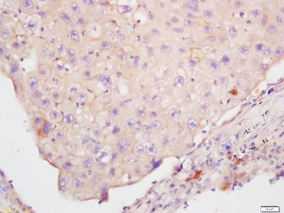 Tissue/cell: human lung carcinoma; 4% Paraformaldehyde-fixed and paraffin-embedded; Tissue/cell: human lung carcinoma; 4% Paraformaldehyde-fixed and paraffin-embedded;Antigen retrieval: citrate buffer ( 0.01M, pH 6.0 ), Boiling bathing for 15min; Block endogenous peroxidase by 3% Hydrogen peroxide for 30min; Blocking buffer (normal goat serum,C-0005) at 37℃ for 20 min; Incubation: Anti-KRAS Polyclonal Antibody, Unconjugated(bs-1033R) 1:200, overnight at 4°C, followed by conjugation to the secondary antibody(SP-0023) and DAB(C-0010) staining 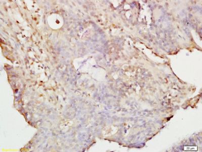 Tissue/cell: rat colon tissue; 4% Paraformaldehyde-fixed and paraffin-embedded; Tissue/cell: rat colon tissue; 4% Paraformaldehyde-fixed and paraffin-embedded;Antigen retrieval: citrate buffer ( 0.01M, pH 6.0 ), Boiling bathing for 15min; Block endogenous peroxidase by 3% Hydrogen peroxide for 30min; Blocking buffer (normal goat serum,C-0005) at 37℃ for 20 min; Incubation: Anti-KRAS Polyclonal Antibody, Unconjugated(bs-1033R) 1:200, overnight at 4°C, followed by conjugation to the secondary antibody(SP-0023) and DAB(C-0010) staining Tissue/cell: human endometrium tissue; 4% Paraformaldehyde-fixed and paraffin-embedded; Antigen retrieval: citrate buffer ( 0.01M, pH 6.0 ), Boiling bathing for 15min; Block endogenous peroxidase by 3% Hydrogen peroxide for 30min; Blocking buffer (normal goat serum,C-0005) at 37℃ for 20 min; Incubation: Anti-KRAS Polyclonal Antibody, Unconjugated(bs-1033R) 1:200, overnight at 4°C, followed by conjugation to the secondary antibody(SP-0023) and DAB(C-0010) staining 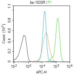 Blank control (Black line):Molt4 (Black). Blank control (Black line):Molt4 (Black).Primary Antibody (green line): Rabbit Anti-KRAS antibody (bs-1033R) Dilution: 1μg /10^6 cells; Isotype Control Antibody (orange line): Rabbit IgG . Secondary Antibody (white blue line): Goat anti-rabbit IgG-AF647 Dilution: 1μg /test. Protocol The cells were fixed with 4% PFA (10min at room temperature)and then permeabilized with 90% ice-cold methanol for 20 min at room temperature. The cells were then incubated in 5%BSA to block non-specific protein-protein interactions for 30 min at room temperature .Cells stained with Primary Antibody for 30 min at room temperature. The secondary antibody used for 40 min at room temperature. Acquisition of 20,000 events was performed. |
我要詢價
*聯系方式:
(可以是QQ、MSN、電子郵箱、電話等,您的聯系方式不會被公開)
*內容:


