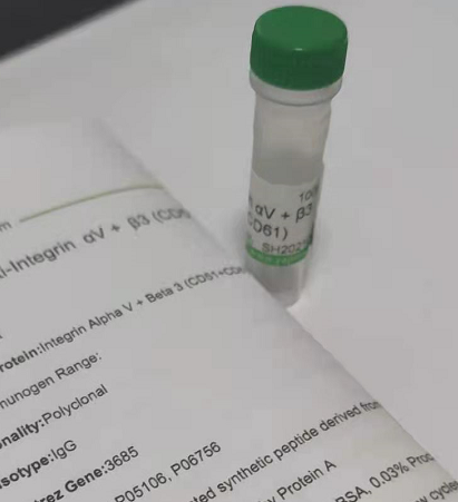
| 中文名稱 | 整合素αVβ3抗體 |
| 別 名 | CD51 + CD61; CD51; CD61; GP3A; GPIIIa; integrin alpha v; integrin beta 3; intregrin alpha v beta 3; ITGAV; ITGB3; Platelet membrane glycoprotein IIIa; Vitronectin receptor alpha subunit; VNRA; Antigen identified by monoclonal antibody L230; CD51 antigen; CD61 antigen; DKFZp686A08142; Integrin alpha V (vitronectin receptor, alpha polypeptide, antigen CD51); integrin alpha v; Integrin beta 3 (platelet glycoprotein IIIa, antigen CD61); Integrin beta chain beta 3; intregrin alpha v beta 3; ITGAV; MSK8; Platelet glycoprotein IIIa; Platelet membrane glycoprotein IIIa; Vitronectin receptor alpha subunit; Vitronectin receptor subunit alpha; avb3. |
| 研究領域 | 細胞生物 免疫學 信號轉導 干細胞 細胞粘附分子 |
| 抗體來源 | Rabbit |
| 克隆類型 | Polyclonal |
| 交叉反應 | Human, Mouse, Rat, |
| 產品應用 | WB=1:500-2000 ELISA=1:500-1000 IHC-P=1:100-500 IHC-F=1:100-500 Flow-Cyt=1μg/Test IF=1:100-500 (石蠟切片需做抗原修復) not yet tested in other applications. optimal dilutions/concentrations should be determined by the end user. |
| 細胞定位 | 細胞膜 |
| 性 狀 | Liquid |
| 濃 度 | 1mg/ml |
| 免 疫 原 | KLH conjugated synthetic peptide derived from human Integrin Alpha V + Beta 3: |
| 亞 型 | IgG |
| 純化方法 | affinity purified by Protein A |
| 儲 存 液 | 0.01M TBS(pH7.4) with 1% BSA, 0.03% Proclin300 and 50% Glycerol. |
| 保存條件 | Shipped at 4℃. Store at -20 °C for one year. Avoid repeated freeze/thaw cycles. |
| PubMed | PubMed |
| 產品介紹 | Integrins are integral cell-surface proteins composed of an alpha chain and a beta chain. They are known to participate in cell adhesion as well as cell-surface mediated signalling. Integrins are heterodimeric integral membrane proteins composed of an alpha chain and a beta chain. CD51 encodes integrin alpha chain V. The I-domain containing integrin alpha V undergoes post-translational cleavage to yield disulfide-linked heavy and light chains, that combine with multiple integrin beta chains to form different integrins. The CD61 protein product is the integrin beta chain beta 3. Integrin beta 3 is found along with the alpha IIb chain in platelets. Integrin alpha V/beta 3 is a receptor for cytotactin, fibronectin, laminin, matrix metalloproteinase 2, osteopontin, osteomodulin, prothrombin, thrombospondin, vitronectin and von Willebrand factor. Integrin alpha V/beta 3 recognizes the sequence R-G-D in a wide array of ligands. The alpha V integrins are receptors for vitronectin, cytotactin, fibronectin, fibrinogen, laminin, matrix metalloproteinase 2, osteopontin, osteomodulin, prothrombin, thrombospondin and von Willebrand factor. They recognize the sequence R-G-D in a wide array of ligands. Subcellular Location: Cell Membrane; single-pass type I membrane protein. SWISS: P05106, P06756 Gene ID: 3685 Database links: Entrez Gene: 3685 Human Entrez Gene: 3690 Human Omim: 173470 Human Omim: 193210 Human SwissProt: P05106 Human SwissProt: P06756 Human Unigene: 218040 Human Unigene: 436873 Human Important Note: This product as supplied is intended for research use only, not for use in human, therapeutic or diagnostic applications. 整合素αVβ3為二聚體的跨膜糖蛋白質,可以調節腫瘤細胞在多種細胞外基質蛋白中的粘附和遷移,在被激活的內皮細胞中有較高的表達,并在新生血管生成過程中發揮優勢。 整合素αVβ3在細胞外的信號傳入細胞內調節細胞生長、改變細胞形態、影響細胞運動, 并在腫瘤侵襲和轉移的過程中起重要作用.整合素αVβ3是特異性表達在血管內皮細胞表面的粘附因子。 |
| 產品圖片 |
(secondary antibody)Goat Anti-rabbit IgG/FITC (bs-0295G-FITC), 1:00, at 4°C for 40 minutes. |



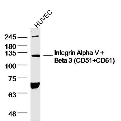 Sample: HUVEC (human)Cell Lysate at 40 ug
 Tissue/cell: Human laryngeal tissue; 4% Paraformaldehyde-fixed and paraffin-embedded;
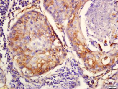 Tissue/cell: Human lung cancer tissue; 4% Paraformaldehyde-fixed and paraffin-embedded;
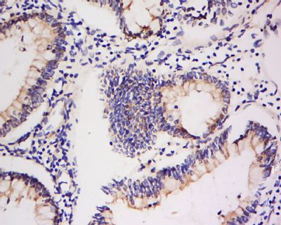 cell: mouse lung.
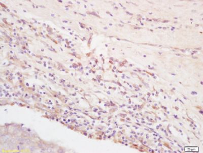 Tissue/cell: rat brain tissue; 4% Paraformaldehyde-fixed and paraffin-embedded;
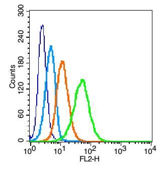 Blank control: U937(blue).
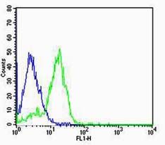 Cell: MCF-7


