| 中文名稱 | 腎小球細胞粘附分子受體抗體 |
| 別 名 | CNF; NPHN; Nephrosis 1 congenital Finnish type; NPHS 1; NPHS1; Renal glomerulus specific cell adhesion receptor; Renal glomerulus-specific cell adhesion receptor; NPHN_HUMAN . |
| 抗體來源 | Rabbit |
| 克隆類型 | Polyclonal |
| 交叉反應 | Human, Mouse, Rat, Dog, (predicted: Rabbit, ) |
| 產(chǎn)品應用 | WB=1:500-2000 ELISA=1:500-1000 IHC-P=1:100-500 Flow-Cyt=1μg/Test (石蠟切片需做抗原修復) not yet tested in other applications. optimal dilutions/concentrations should be determined by the end user. |
| 分 子 量 | 138kDa |
| 細胞定位 | 細胞外基質(zhì) |
| 性 狀 | Liquid |
| 濃 度 | 1mg/ml |
| 免 疫 原 | KLH conjugated synthetic peptide derived from human Nephrin:451-550/1241 |
| 亞 型 | IgG |
| 純化方法 | affinity purified by Protein A |
| 儲 存 液 | 0.01M TBS(pH7.4) with 1% BSA, 0.03% Proclin300 and 50% Glycerol. |
| 保存條件 | Shipped at 4℃. Store at -20 °C for one year. Avoid repeated freeze/thaw cycles. |
| PubMed | PubMed |
| 產(chǎn)品介紹 | This gene encodes a member of the immunoglobulin family of cell adhesion molecules that functions in the glomerular filtration barrier in the kidney. The gene is primarily expressed in renal tissues, and the protein is a type-1 transmembrane protein found at the slit diaphragm of glomerular podocytes. The slit diaphragm is thought to function as an ultrafilter to exclude albumin and other plasma macromolecules in the formation of urine. Mutations in this gene result in Finnish-type congenital nephrosis 1, characterized by severe proteinuria and loss of the slit diaphragm and foot processes.[provided by RefSeq, Oct 2009] Function: Seems to play a role in the development or function of the kidney glomerular filtration barrier. Regulates glomerular vascular permeability. May anchor the podocyte slit diaphragm to the actin cytoskeleton. Plays a role in skeletal muscle formation through regulation of myoblast fusion. Subunit: Interacts with CD2AP (via C-terminal domain). Interacts with MAGI1 (via PDZ 2 and 3 domains) forming a tripartite complex with IGSF5/JAM4. Interacts with DDN; the interaction is direct. Self-associates (via the Ig-like domains). Also interacts (via the Ig-like domains) with KIRREL/NEPH1 and KIRREL2; the interaction with KIRREL is dependent on KIRREL glycosylation. Forms a complex with ACTN4, CASK, IQGAP1, MAGI2, SPTAN1 and SPTBN1 (By similarity). Interacts with NPHS2. Subcellular Location: Cell membrane; Single-pass type I membrane protein (Potential). Note=Predominantly located at podocyte slit diaphragm between podocyte foot processes. Also associated with podocyte apical plasma membrane. Tissue Specificity: Specifically expressed in podocytes of kidney glomeruli. Expressed in kidney glomeruli. In the embryo,expressed in the mesonephric kidney at E11 with strong expression in cranial tubules with podocyte-like structures. Expression is observed in the podocytes of the developing kidney from E13. High expression is also detected in the developing cerebellum, hindbrain, spinal cord, retina and hypothalamus. Expressed in skeletal muscle during myoblast fusion such as in the adult following acute injury and in the embryo but not detected in uninjured adult skeletal muscle. Isoform 1 and isoform 2 are expressed in the newborn brain and developing cerebellum. Isoform 1 is the predominant isoform in adult kidney Post-translational modifications: Phosphorylated at Tyr-1193 by FYN, leading to the recruitment and activation of phospholipase C-gamma-1/PLCG1. DISEASE: Defects in NPHS1 are the cause of nephrotic syndrome type 1 (NPHS1) [MIM:256300]; also known as Finnish congenital nephrosis (CNF). A renal disease characterized clinically by proteinuria, hypoalbuminemia, hyperlipidemia, and edema. Kidney biopsies show non-specific histologic changes such as focal segmental glomerulosclerosis and diffuse mesangial proliferation. Some affected individuals have an inherited steroid-resistant form and progress to end-stage renal failure. Similarity: Belongs to the immunoglobulin superfamily. Contains 1 fibronectin type-III domain. Contains 8 Ig-like C2-type (immunoglobulin-like) domains. SWISS: O60500 Gene ID: 4868 Database links: Entrez Gene: 4868 Human Entrez Gene: 54631 Mouse Entrez Gene: 64563 Rat Omim: 602716 Human SwissProt: O60500 Human SwissProt: Q9QZS7 Mouse SwissProt: Q9R044 Rat Unigene: 122186 Human Unigene: 437830 Mouse Unigene: 48745 Rat
Important Note: This product as supplied is intended for research use only, not for use in human, therapeutic or diagnostic applications. 葛博:bs-10233R 細胞定位:質(zhì)膜、細胞外基質(zhì) 17.10.19日張鳳英修改 |
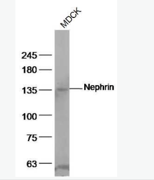
| 產(chǎn)品圖片 | 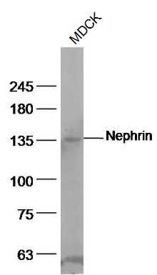 Sample: Sample:MDCK(Dog) Cell Lysate at 40 ug Primary: Anti-Nephrin (bs-10233R) at 1/500 dilution Secondary: IRDye800CW Goat Anti-Rabbit IgG at 1/20000 dilution Predicted band size: 138 kD Observed band size: 138 kD 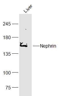 Sample: Sample:Liver (Mouse) Lysate at 40 ug Primary: Anti-Nephrin (bs-10233R) at 1/300 dilution Secondary: IRDye800CW Goat Anti-Rabbit IgG at 1/20000 dilution Predicted band size: 138 kD Observed band size: 138 kD 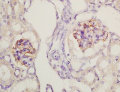 Tissue/cell: mouse kidney tissue; 4% Paraformaldehyde-fixed and paraffin-embedded; Tissue/cell: mouse kidney tissue; 4% Paraformaldehyde-fixed and paraffin-embedded;Antigen retrieval: citrate buffer ( 0.01M, pH 6.0 ), Boiling bathing for 15min; Block endogenous peroxidase by 3% Hydrogen peroxide for 30min; Blocking buffer (normal goat serum,C-0005) at 37℃ for 20 min; Incubation: Anti-Nephrin Polyclonal Antibody, Unconjugated(bs-10233R) 1:200, overnight at 4°C, followed by conjugation to the secondary antibody(SP-0023) and DAB(C-0010) staining 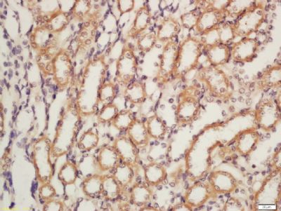 Tissue/cell: rat kidney tissue; 4% Paraformaldehyde-fixed and paraffin-embedded; Tissue/cell: rat kidney tissue; 4% Paraformaldehyde-fixed and paraffin-embedded;Antigen retrieval: citrate buffer ( 0.01M, pH 6.0 ), Boiling bathing for 15min; Block endogenous peroxidase by 3% Hydrogen peroxide for 30min; Blocking buffer (normal goat serum,C-0005) at 37℃ for 20 min; Incubation: Anti-Nephrin Polyclonal Antibody, Unconjugated(bs-10233R) 1:200, overnight at 4°C, followed by conjugation to the secondary antibody(SP-0023) and DAB(C-0010) staining 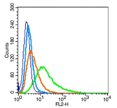 Blank control: RSC96(blue), the cells were fixed with 2% paraformaldehyde (10 min) and then permeabilized with ice-cold 90% methanol for 30 min on ice. Blank control: RSC96(blue), the cells were fixed with 2% paraformaldehyde (10 min) and then permeabilized with ice-cold 90% methanol for 30 min on ice.Isotype Control Antibody: Rabbit IgG(orange) ; Secondary Antibody: Goat anti-rabbit IgG-PE(white blue), Dilution: 1:200 in 1 X PBS containing 0.5% BSA ; Primary Antibody Dilution: 1μg in 100 μL1X PBS containing 0.5% BSA(green).  the cells(293T) were fixed with 2% paraformaldehyde (10 min). the cells(293T) were fixed with 2% paraformaldehyde (10 min).Isotype Control Antibody: Rabbit IgG(orange) ; Secondary Antibody: Goat anti-rabbit IgG-PE(white blue), Dilution: 1:200 in 1 X PBS containing 0.5% BSA ; Primary Antibody Dilution: 1μg in 100 μL1X PBS containing 0.5% BSA(green). |
我要詢價
*聯(lián)系方式:
(可以是QQ、MSN、電子郵箱、電話等,您的聯(lián)系方式不會被公開)
*內(nèi)容:


