| 中文名稱 | CD19抗體 |
| 別 名 | Antibody deficiency due to defect in CD19, included; AW495831; B lymphocyte antigen CD19; B lymphocyte surface antigen B4; B4; CD 19; CD19 antigen; CD19 molecule; Cd19 protein; Differentiation Antigen CD19; Leu 12; Leu12; Lymphocyte Surface Antigen; MGC109570; MGC12802; T-cell surface antigen Leu-12; CD19_HUMAN. |
| 研究領域 | 細胞生物 免疫學 干細胞 細胞表面分子 淋巴細胞 b-淋巴細胞 |
| 抗體來源 | Rabbit |
| 克隆類型 | Polyclonal |
| 交叉反應 | Human, Mouse, Rat, (predicted: Pig, Cow, Horse, Guinea Pig, ) |
| 產品應用 | ELISA=1:500-1000 IHC-P=1:100-500 IHC-F=1:100-500 Flow-Cyt=1μg/Test IF=1:100-500 (石蠟切片需做抗原修復) not yet tested in other applications. optimal dilutions/concentrations should be determined by the end user. |
| 分 子 量 | 59kDa |
| 細胞定位 | 細胞膜 |
| 性 狀 | Liquid |
| 濃 度 | 1mg/ml |
| 免 疫 原 | KLH conjugated synthetic peptide derived from human CD19:485-556/556 |
| 亞 型 | IgG |
| 純化方法 | affinity purified by Protein A |
| 儲 存 液 | 0.01M TBS(pH7.4) with 1% BSA, 0.03% Proclin300 and 50% Glycerol. |
| 保存條件 | Shipped at 4℃. Store at -20 °C for one year. Avoid repeated freeze/thaw cycles. |
| PubMed | PubMed |
| 產品介紹 | CD19 is a transmembrane glycoprotein that is delectively expressed on the cell surface of B-lymphocytes,where it activates intracellular signaling cascades involving both Ras and phosphatidylinositol 3-kinase pathways.Lymphocytes proliferate and differentiate in response to various concentrations of different antigens. The ability of the B cell to respond in a specific, yet sensitive manner to the various antigens is achieved with the use of low-affinity antigen receptors. This gene encodes a cell surface molecule which assembles with the antigen receptor of B lymphocytes in order to decrease the threshold for antigen receptor-dependent stimulation. Function: Assembles with the antigen receptor of B-lymphocytes in order to decrease the threshold for antigen receptor-dependent stimulation. Subunit: Forms a complex with CD21, CD81 and CD225 in the membrane of mature B-cells. Interacts with VAV. Interacts with GRB2 and SOS when phosphorylated on Tyr-348 and/or Tyr-378. Interacts with PLCG2 when phosphorylated on Tyr-409. Interacts with LYN. Subcellular Location: Membrane; Single-pass type I membrane protein. Post-translational modifications: Phosphorylated on serine and threonine upon DNA damage, probably by ATM or ATR. Phosphorylated on tyrosine following B-cell activation. Phosphorylated on tyrosine residues by LYN. DISEASE: Defects in CD19 are the cause of immunodeficiency common variable type 3 (CVID3) [MIM:613493]; also called antibody deficiency due to CD19 defect. CVID3 is a primary immunodeficiency characterized by antibody deficiency, hypogammaglobulinemia, recurrent bacterial infections and an inability to mount an antibody response to antigen. The defect results from a failure of B-cell differentiation and impaired secretion of immunoglobulins; the numbers of circulating B-cells is usually in the normal range, but can be low. Similarity: Contains 2 Ig-like C2-type (immunoglobulin-like) domains. SWISS: P15391 Gene ID: 930 Database links: Entrez Gene: 930 Human Entrez Gene: 12478 Mouse Entrez Gene: 365367 Rat Omim: 107265 Human SwissProt: P15391 Human SwissProt: P25918 Mouse Unigene: 652262 Human Unigene: 4360 Mouse Important Note: This product as supplied is intended for research use only, not for use in human, therapeutic or diagnostic applications. CD19是一種質膜蛋白質,參與信號傳導作用。表達與前B細胞和成熟的B細胞膜表面,與B細胞的活化調節和發育調節相關,在T細胞和正常粒細胞上無表達。此抗體可以特異性識別CD19,主要用于標記正常B細胞及腫瘤性B細胞。 |
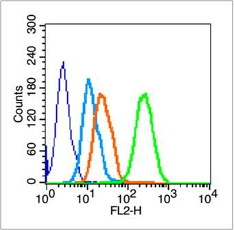
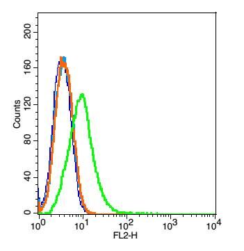
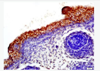
| 產品圖片 |  Tissue/cell: mouse fetal skin; 4% Paraformaldehyde-fixed and paraffin-embedded; Tissue/cell: mouse fetal skin; 4% Paraformaldehyde-fixed and paraffin-embedded;Antigen retrieval: citrate buffer ( 0.01M, pH 6.0 ), Boiling bathing for 15min; Block endogenous peroxidase by 3% Hydrogen peroxide for 30min; Blocking buffer (normal goat serum,C-0005) at 37℃ for 20 min; Incubation: Anti-CD19 Polyclonal Antibody, Unconjugated(bs-0079R) 1:500, overnight at 4°C, followed by conjugation to the secondary antibody(SP-0023) and DAB(C-0010) staining   Blank control (blue line): HL60 cells (blue). Blank control (blue line): HL60 cells (blue).Primary Antibody (green line): Rabbit Anti-CD19 antibody (bs-0079R) Dilution: 1μg /10^6 cells; Isotype Control Antibody (orange line): Rabbit IgG . Secondary Antibody (white blue line): Goat anti-rabbit IgG-PE Dilution: 1μg /test. Protocol The cells were fixed with 70% methanol (Overnight at 4℃) . Cells stained with Primary Antibody for 30 min at room temperature. The cells were then incubated in 1 X PBS/2%BSA/10% goat serum to block non-specific protein-protein interactions followed by the antibody for 15 min at room temperature. The secondary antibody used for 40 min at room temperature. Acquisition of 20,000 events was performed.  Blank control: Raji(blue). Blank control: Raji(blue).Primary Antibody: Rabbit Anti-CD19 antibody(bs-0079R), Dilution: 5μg in 100 μL 1X PBS containing 0.5% BSA; Isotype Control Antibody: Rabbit IgG (orange) ,used under the same conditions. Secondary Antibody: Goat anti-rabbit IgG-PE(white blue), Dilution: 1:200 in 1 X PBS containing 0.5% BSA. Protocol Primary antibody (bs-0079R, 5μg /1x10^6 cells) were incubated for 30 min on the ice, followed by 1 X PBS containing 0.5% BSA + 1 0% goat serum (15 min) to block non-specific protein-protein interactions. Then the Goat Anti-rabbit IgG/PE antibody was added into the blocking buffer mentioned above to react with the primary antibody at 1/200 dilution for 30 min on ice. Acquisition of 20,000 events was performed. |
我要詢價
*聯系方式:
(可以是QQ、MSN、電子郵箱、電話等,您的聯系方式不會被公開)
*內容:


