| 中文名稱 | FC段γ受體3/免疫球蛋白G Fc段受體III抗體 |
| 別 名 | FCG3A_HUMAN; CD 16; CD 16a; CD16A; CD16a antigen; CD16B; CD16b antigen; Fc fragment of IgG; Fc fragment of IgG low affinity IIIa receptor (CD16); Fc fragment of IgG, low affinity III, receptor (CD16); Fc fragment of IgG, low affinity IIIa, receptor (CD16); Fc fragment of IgG, low affinity IIIa, receptor (CD16a); Fc fragment of IgG, low affinity IIIb, receptor (CD16b); Fc fragment of IgG, low affinity IIIb, receptor for (CD16); Fc gamma R3; Fc gamma receptor IIIA; Fc gamma receptor IIIb (CD 16); Fc gamma RIII alpha; Fc gamma RIII; Fc gamma RIII beta; Fc gamma RIIIa; Fc gamma RIIIb; Fc of IgG; Fc-gamma receptor III2 (CD 16); Fc-gamma receptor III2 (CD16); Fc-gamma receptor IIIb (CD16); FCG 3; FCG3; FCgammaRIIIA; FCGR 3; FCGR 3A; FCGR3; FCGR3A; FCGR3A protein; FCGRIII; FcR 10; FcR10; FcRIII; FcRIIIa; IGFR 3; IGFR3; IgG Fc receptor III 1; IgG Fc receptor III 2; immunoglobulin G Fc receptor III; Low affinity IIIa receptor; Low affinity immunoglobulin gamma Fc region receptor III A; Low affinity immunoglobulin gamma Fc region receptor IIIB; neutrophil-specific antigen NA. |
| 研究領域 | 細胞生物 免疫學 干細胞 細胞表面分子 細胞類型標志物 |
| 抗體來源 | Rabbit |
| 克隆類型 | Polyclonal |
| 交叉反應 | Human, Mouse, Rat, (predicted: Pig, Cow, Rabbit, Sheep, ) |
| 產品應用 | WB=1:500-2000 ELISA=1:500-1000 IHC-P=1:100-500 IHC-F=1:100-500 Flow-Cyt=1ug/Test IF=1:100-500 (石蠟切片需做抗原修復) not yet tested in other applications. optimal dilutions/concentrations should be determined by the end user. |
| 分 子 量 | 27kDa |
| 細胞定位 | 細胞膜 分泌型蛋白 |
| 性 狀 | Liquid |
| 濃 度 | 1mg/ml |
| 免 疫 原 | KLH conjugated synthetic peptide derived from human IGFR3/CD16:131-230/254 |
| 亞 型 | IgG |
| 純化方法 | affinity purified by Protein A |
| 儲 存 液 | 0.01M TBS(pH7.4) with 1% BSA, 0.03% Proclin300 and 50% Glycerol. |
| 保存條件 | Shipped at 4℃. Store at -20 °C for one year. Avoid repeated freeze/thaw cycles. |
| PubMed | PubMed |
| 產品介紹 | This gene encodes a receptor for the Fc portion of immunoglobulin G, and it is involved in the removal of antigen-antibody complexes from the circulation, as well as other other antibody-dependent responses. This gene (FCGR3A) is highly similar to another nearby gene (FCGR3B) located on chromosome 1. The receptor encoded by this gene is expressed on natural killer(NK) cells as an integral membrane glycoprotein anchored through a transmembrane peptide, whereas FCGR3B is expressed on polymorphonuclear neutrophils (PMN) where the receptor is anchored through a phosphatidylinositol (PI) linkage. Mutations in this gene have been linked to susceptibility to recurrent viral infections, susceptibility to systemic lupus erythematosus, and alloimmune neonatal neutropenia. Alternatively spliced transcript variants encoding different isoforms have been found for this gene. [provided by RefSeq, Jul 2008]. Function: Receptor for the Fc region of IgG. Binds complexed or aggregated IgG and also monomeric IgG. Mediates antibody-dependent cellular cytotoxicity (ADCC) and other antibody-dependent responses, such as phagocytosis. Subunit: Exists as a heterooligomeric receptor complex with Fc epsilon receptor I gamma subunit and / or the CD3 zeta subunit. Interacts with INPP5D/SHIP1. Subcellular Location: Cell membrane. Secreted. Exists also as a soluble receptor. Tissue Specificity: Expressed on natural killer cells, macrophages, subpopulation of T-cells, immature thymocytes and placental trophoblasts. Post-translational modifications: Glycosylated. Contains high mannose- and complex-type oligosaccharides. The soluble form is produced by a proteolytic cleavage. [MISCELLANEOUS] Encoded by one of two nearly indentical genes: FCGR3A (Shown here) and FCGR3B which are expressed in a tissue-specific manner. The Phe-203 in III-A determines the transmembrane domains whereas the 'Ser-203' in III-B determines the GPI-anchoring. Similarity: Contains 2 Ig-like C2-type (immunoglobulin-like) domains. SWISS: P08637 Gene ID: 2214 Database links: Entrez Gene: 2214 Human Entrez Gene: 14131 Mouse Entrez Gene: 304966 Rat Omim: 146740 Human SwissProt: P08637 Human SwissProt: P08508 Mouse SwissProt: P27645 Rat Unigene: 372679 Human Important Note: This product as supplied is intended for research use only, not for use in human, therapeutic or diagnostic applications. |
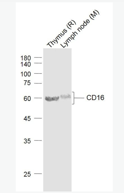
| 產品圖片 | 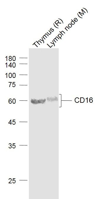 Sample: Sample:Lane 1: Thymus (Rat) Lysate at 40 ug Lane 2: Lymph node (Mouse) Lysate at 40 ug Primary: Anti-CD16 (bs-6028R) at 1/1000 dilution Secondary: IRDye800CW Goat Anti-Rabbit IgG at 1/20000 dilution Predicted band size: 55 kD Observed band size: 60 kD 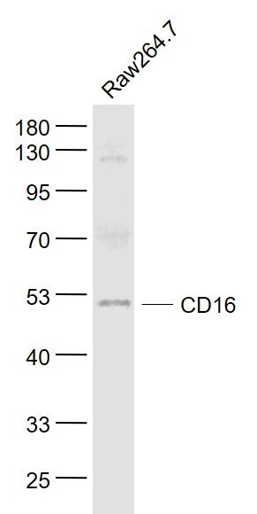 Sample: Sample:Raw264.7(Mouse) Cell Lysate at 30 ug Primary: Anti- CD16 (bs-6028R) at 1/1000 dilution Secondary: IRDye800CW Goat Anti-Rabbit IgG at 1/20000 dilution Predicted band size: 27 kD Observed band size: 50 kD 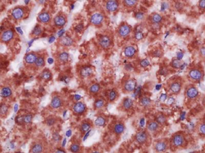 Paraformaldehyde-fixed, paraffin embedded (Rat liver); Antigen retrieval by boiling in sodium citrate buffer (pH6.0) for 15min; Block endogenous peroxidase by 3% hydrogen peroxide for 20 minutes; Blocking buffer (normal goat serum) at 37°C for 30min; Antibody incubation with (CD16) Polyclonal Antibody, Unconjugated (bs-6028R) at 1:400 overnight at 4°C, followed by operating according to SP Kit(Rabbit) (sp-0023) instructionsand DAB staining. Paraformaldehyde-fixed, paraffin embedded (Rat liver); Antigen retrieval by boiling in sodium citrate buffer (pH6.0) for 15min; Block endogenous peroxidase by 3% hydrogen peroxide for 20 minutes; Blocking buffer (normal goat serum) at 37°C for 30min; Antibody incubation with (CD16) Polyclonal Antibody, Unconjugated (bs-6028R) at 1:400 overnight at 4°C, followed by operating according to SP Kit(Rabbit) (sp-0023) instructionsand DAB staining.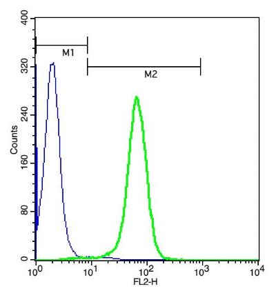 Cell: U937(4% Paraformaldehyde fixed for 10 minutes,2% BSA at 4∑blocked for 30 minutes.). Cell: U937(4% Paraformaldehyde fixed for 10 minutes,2% BSA at 4∑blocked for 30 minutes.).Concentration:1:100;Incubation: 40 minutes. Flow cytometric analysis of Rabbit Anti-CD16 antibody (bs-6028R)(green) compared with control in the absence of primary antibody (blue) followed by U937. Secondary antibody: Goat Anti-rabbit IgG/PE antibody (bs-0295G-PE) |
我要詢價
*聯系方式:
(可以是QQ、MSN、電子郵箱、電話等,您的聯系方式不會被公開)
*內容:


