| 中文名稱 | 自噬微管相關蛋白輕鏈3抗體 |
| 別 名 | Autophagy related protein LC3 A; Autophagy related protein LC3 B; Autophagy related ubiquitin like modifier LC3 A; Autophagy related ubiquitin like modifier LC3 B; MAP1 light chain 3 like protein 1; MAP1 light chain 3 like protein 2; MAP1A/1B light chain 3 A; MAP1A/1B light chain 3 B; MAP1A/1BLC3; MAP1A/MAP1B LC3 A; MAP1A/MAP1B LC3 B; MAP1ALC3; MAP1BLC3; MAP1LC3A; MAP1LC3B; Microtubule associated protein 1 light chain 3 alpha; Microtubule associated protein 1 light chain 3 beta; Microtubule associated proteins 1A/1B light chain 3; Microtubule associated proteins 1A/1B light chain 3A; Microtubule associated proteins 1A/1B light chain 3B; MLP3A_HUMAN. |
| 研究領域 | 腫瘤 細胞生物 神經生物學 信號轉導 細胞骨架 細胞外基質 細胞自噬 |
| 抗體來源 | Rabbit |
| 克隆類型 | Polyclonal |
| 交叉反應 | Human, Rat, (predicted: Mouse, Chicken, Dog, Pig, Cow, Horse, Sheep, Goat, ) |
| 產品應用 | WB=1:500-2000 ELISA=1:500-1000 IHC-P=1:100-500 IHC-F=1:100-500 Flow-Cyt=1μg/Test ICC=1:100-500 IF=1:100-500 (石蠟切片需做抗原修復) not yet tested in other applications. optimal dilutions/concentrations should be determined by the end user. |
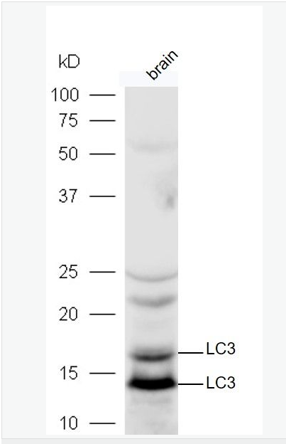
| 分 子 量 | 14/16kDa |
| 細胞定位 | 細胞漿 |
| 性 狀 | Liquid |
| 濃 度 | 1mg/ml |
| 免 疫 原 | KLH conjugated synthetic peptide derived from human LC3:31-121/121 |
| 亞 型 | IgG |
| 純化方法 | affinity purified by Protein A |
| 儲 存 液 | 0.01M TBS(pH7.4) with 1% BSA, 0.03% Proclin300 and 50% Glycerol. |
| 保存條件 | Shipped at 4℃. Store at -20 °C for one year. Avoid repeated freeze/thaw cycles. |
| PubMed | PubMed |
| 產品介紹 | A major contributor to cellular homeostasis is the ability of the cell to strike a balance between the formation and degradation/removal of its cellular components. This process of internal cellular turn-over is called autophagy (self-eating), and is facilitated by a pathway of around 16 interacting proteins in the human. LC3, a ubiquitin-like modifier protein, is the human homolog of yeast Apg8 and is involved in the formation of autophagosomal vacuoles, called autophagosomes. LC3 is expressed as 3 splice variants (LC3A, LC3B and LC3C), which exhibit different tissue distributions and are processed into cytosolic and autophagosomal membrane-bound forms, termed LC3-I and LC3-II, respectively. A disruption to the autophagic process is now associated with the progression of several cancers, neurodegenerative disorders and cardiac pathologies, where LC3 is widely employed as a marker for autophagy. Function: Probably involved in formation of autophagosomal vacuoles (autophagosomes). Subunit: 3 different light chains, LC1, LC2 and LC3, can associate with MAP1A and MAP1B proteins. Interacts with SQSTM1. Interacts with TP53INP1 and TP53INP2. Subcellular Location: Cytoplasmic. Endomembrane system; Lipid-anchor. Cytoplasmic vesicle, autophagosome membrane; Lipid-anchor. Note: LC3B binds to the autophagic membranes. Tissue Specificity: Most abundant in heart, brain, liver, skeletal muscle and testis but absent in thymus and peripheral blood leukocytes. Post-translational modifications: The precursor molecule is cleaved by APG4B/ATG4B to form the cytosolic form, LC3-I. This is activated by APG7L/ATG7, transferred to ATG3 and conjugated to phospholipid to form the membrane-bound form, LC3-II. Similarity: Belongs to the MAP1 LC3 family. SWISS: Q9H492 Gene ID: 84557 Database links: Entrez Gene: 84557 Human Entrez Gene: 66734 Mouse Entrez Gene: 362245 Rat Omim: 601242 Human SwissProt: Q9H492 Human SwissProt: Q91VR7 Mouse SwissProt: Q6XVN8 Rat Unigene: 632273 Human Unigene: 196239 Mouse Unigene: 3135 Rat Important Note: This product as supplied is intended for research use only, not for use in human, therapeutic or diagnostic applications. |
| 產品圖片 | 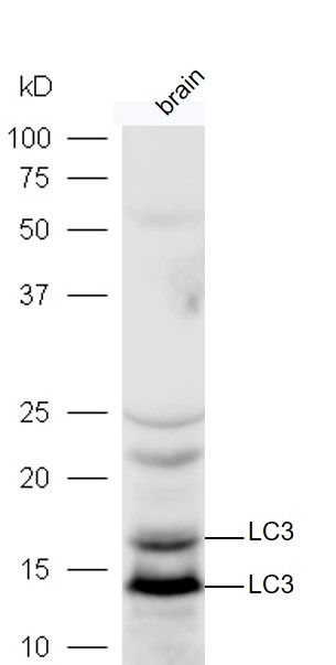 Protein: mouse brain lysates at 40ug; Protein: mouse brain lysates at 40ug;Primary: rabbit Anti-LC3 (bs-8878R) at 1:300; Secondary: HRP conjugated Goat-Anti-rabbit IgG(bs-0295G-HRP) at 1: 5000; Predicted band size:14 kD Observed band size:14,16 kD 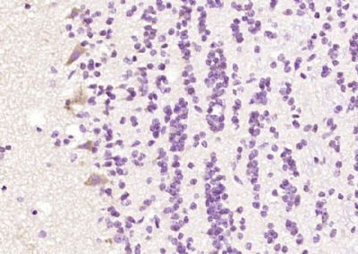 Paraformaldehyde-fixed, paraffin embedded (rat brain); Antigen retrieval by boiling in sodium citrate buffer (pH6.0) for 15min; Block endogenous peroxidase by 3% hydrogen peroxide for 20 minutes; Blocking buffer (normal goat serum) at 37°C for 30min; Antibody incubation with (LC3) Polyclonal Antibody, Unconjugated (bs-8878R) at 1:200 overnight at 4°C, followed by operating according to SP Kit(Rabbit) (sp-0023) instructionsand DAB staining. Paraformaldehyde-fixed, paraffin embedded (rat brain); Antigen retrieval by boiling in sodium citrate buffer (pH6.0) for 15min; Block endogenous peroxidase by 3% hydrogen peroxide for 20 minutes; Blocking buffer (normal goat serum) at 37°C for 30min; Antibody incubation with (LC3) Polyclonal Antibody, Unconjugated (bs-8878R) at 1:200 overnight at 4°C, followed by operating according to SP Kit(Rabbit) (sp-0023) instructionsand DAB staining.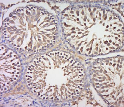 Paraformaldehyde-fixed, paraffin embedded (rat testis tissue); Antigen retrieval by boiling in sodium citrate buffer (pH6.0) for 15min; Block endogenous peroxidase by 3% hydrogen peroxide for 20 minutes; Blocking buffer (normal goat serum) at 37°C for 30min; Antibody incubation with (LC3) Polyclonal Antibody, Unconjugated (bs-8878R) at 1:400 overnight at 4°C, followed by a conjugated secondary (sp-0023) for 20 minutes and DAB staining. Paraformaldehyde-fixed, paraffin embedded (rat testis tissue); Antigen retrieval by boiling in sodium citrate buffer (pH6.0) for 15min; Block endogenous peroxidase by 3% hydrogen peroxide for 20 minutes; Blocking buffer (normal goat serum) at 37°C for 30min; Antibody incubation with (LC3) Polyclonal Antibody, Unconjugated (bs-8878R) at 1:400 overnight at 4°C, followed by a conjugated secondary (sp-0023) for 20 minutes and DAB staining.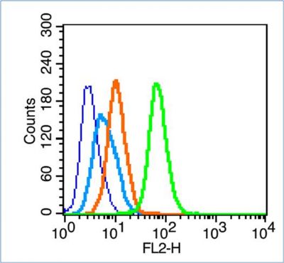 Blank control (blue line): Hela (fixed with 70% ethanol (Overmight at 4℃) and then permeabilized with 90% ice-cold methanol for 30 min at -20℃). Blank control (blue line): Hela (fixed with 70% ethanol (Overmight at 4℃) and then permeabilized with 90% ice-cold methanol for 30 min at -20℃).Primary Antibody (green line): Rabbit Anti-LC3 antibody (bs-8878R),Dilution: 1μg /10^6 cells; Isotype Control Antibody (orange line): Rabbit IgG . Secondary Antibody (white blue line): Goat anti-rabbit IgG-PE,Dilution: 1μg /test. |
我要詢價
*聯系方式:
(可以是QQ、MSN、電子郵箱、電話等,您的聯系方式不會被公開)
*內容:


