| 產品編號 | 17478R |
| 英文名稱 | Melanoma gp100 |
| 中文名稱 | 黑色素瘤相關抗原gp100抗體 |
| 別 名 | 95 kDa melanocyte specific secreted glycoprotein; 95 kDa melanocyte-specific secreted glycoprotein; D12S53E; gp100; M-beta; ME20; ME20 M/ME20 S; ME20-M; ME20-S; ME20M; ME20M/ME20S; ME20S; Melanocyte lineage specific antigen GP100; Melanocyte protein mel 17; Melanocyte protein Pmel 17; Melanocyte protein Pmel 17 precursor; Melanocytes lineage-specific antigen GP100; Melanoma associated ME20 antigen; Melanoma gp100; Melanoma-associated ME20 antigen; Melanosomal matrix protein 17; Melanosomal matrix protein17; P1; p100; p26; PMEL 17; PMEL; PMEL_HUMAN; PMEL17; Premelanosome protein; Secreted melanoma-associated ME20 antigen; SI; SIL; SILV; Silver (mouse homolog) like; Silver homolog; Silver locus protein homolog; Silver, mouse, homolog of. 研究領域 |
| 抗體來源 | Rabbit |
| 克隆類型 | Polyclonal |
| 交叉反應 | Human, (predicted: Horse, ) |
| 產品應用 | WB=1:500-2000 ELISA=1:5000-10000 IHC-P=1:100-500 IHC-F=1:100-500 ICC=1:100-500 IF=1:100-500 (石蠟切片需做抗原修復) not yet tested in other applications. optimal dilutions/concentrations should be determined by the end user. |
| 分 子 量 | 68kDa |
| 細胞定位 | 細胞外基質 分泌型蛋白 |
| 性 狀 | Liquid |
| 濃 度 | 1mg/ml |
| 免 疫 原 | KLH conjugated synthetic peptide derived from human Melanoma gp100:21-120/661 |
| 亞 型 | IgG |
| 純化方法 | affinity purified by Protein A |
| 儲 存 液 | 0.01M TBS(pH7.4) with 1% BSA, 0.03% Proclin300 and 50% Glycerol. |
| 保存條件 | Shipped at 4℃. Store at -20 °C for one year. Avoid repeated freeze/thaw cycles. |
| PubMed | PubMed |
| 產品介紹 | This gene encodes a melanocyte-specific type I transmembrane glycoprotein. The encoded protein is enriched in melanosomes, which are the melanin-producing organelles in melanocytes, and plays an essential role in the structural organization of premelanosomes. This protein is involved in generating internal matrix fibers that define the transition from Stage I to Stage II melanosomes. This protein undergoes a complex pattern of prosttranslational processing and modification that is essential to the proper functioning of the protein. A secreted form of this protein that is released by proteolytic ectodomain shedding may be used as a melanoma-specific serum marker. Alternate splicing results in multiple transcript variants. [provided by RefSeq, Jan 2011] Function: Plays a central role in the biogenesis of melanosomes. Involved in the maturation of melanosomes from stage I to II. The transition from stage I melanosomes to stage II melanosomes involves an elongation of the vesicle, and the appearance within of distinct fibrillar structures. Release of the soluble form, ME20-S, could protect tumor cells from antibody mediated immunity. Subcellular Location: Secreted and Endoplasmic reticulum membrane. Golgi apparatus. Melanosome. Endosome > multivesicular body. Identified by mass spectrometry in melanosome fractions from stage I to stage IV. Localizes predominantly to intralumenal vesicles (ILVs) within multivesicular bodies. Associates with ILVs found within the lumen of premelanosomes and melanosomes and particularly in compartments that serve as precursors to the striated stage II premelanosomes.Target information above from: UniProt accession P40967 The UniProt Consortium The Universal Protein Resource (UniProt) in 2010 Nucleic Acids Res. 38:D142-D148 (2010) . Tissue Specificity: Preferentially expressed in melanomas. Some expression was found in dysplastic nevi. Not found in normal tissues nor in carcinomas. Normally expressed at low levels in quiescent adult melanocytes but overexpressed by proliferating neonatal melanocytes and during tumor growth. Post-translational modifications: A small amount of P1/P100 (major form) undergoes glycosylation to yield P2/P120 (minor form). P2 is cleaved by a furin-like proprotein convertase (PC) in a pH-dependent manner in a post-Golgi, prelysosomal compartment into two disulfide-linked subunits: a large lumenal subunit, M-alpha/ME20-S, and an integral membrane subunit, M-beta. Despite cleavage, only a small fraction of M-alpha is secreted, whereas most M-alpha and M-beta remain associated with each other intracellularly. M-alpha is further processed to M-alpha N and M-alpha C. M-alpha C further undergoes processing to yield M-alpha C1 and M-alpha C3 (M-alpha C2 in the case of PMEL17-is or PMEL17-ls). Formation of intralumenal fibrils in the melanosomes requires the formation of M-alpha that becomes incorporated into the fibrils. Stage II melanosomes harbor only Golgi-modified Pmel17 fragments that are derived from M-alpha and that bear sialylated O-linked oligosaccharides. N-glycosylated. O-glycosylated; contains sialic acid. Similarity: Belongs to the PMEL/NMB family. Contains 1 PKD domain. SWISS: P40967 Gene ID: 6490 Database links: Entrez Gene: 6490 Human Entrez Gene: 20431 Mouse Entrez Gene: 594851 Pig Entrez Gene: 362818 Rat Omim: 155550 Human SwissProt: Q06154 Cow SwissProt: P40967 Human SwissProt: Q60696 Mouse Unigene: 95972 Human Unigene: 16756 Mouse Unigene: 162910 Rat Important Note: This product as supplied is intended for research use only, not for use in human, therapeutic or diagnostic applications. |
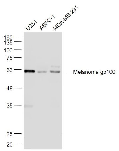
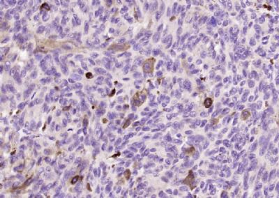
| 產品圖片 |  Sample: U251(Human) Cell Lysate at 30 ug ASPC-1(Human) Cell Lysate at 30 ug MDA-MB-231(Human) Cell Lysate at 30 ug Primary: Anti- Melanoma gp100 (17478R) at 1/1000 dilution Secondary: IRDye800CW Goat Anti-Rabbit IgG at 1/20000 dilution Predicted band size: 68 kD Observed band size: 62 kD  Paraformaldehyde-fixed, paraffin embedded (Human melanoma); Antigen retrieval by boiling in sodium citrate buffer (pH6.0) for 15min; Block endogenous peroxidase by 3% hydrogen peroxide for 20 minutes; Blocking buffer (normal goat serum) at 37°C for 30min; Antibody incubation with (Melanoma gp100) Polyclonal Antibody, Unconjugated (17478R) at 1:200 overnight at 4°C, followed by operating according to SP Kit(Rabbit) (0023) instructionsand DAB staining. |
我要詢價
*聯系方式:
(可以是QQ、MSN、電子郵箱、電話等,您的聯系方式不會被公開)
*內容:


