| 產品編號 | bsm-52290R |
| 英文名稱 | Rabbit Anti-beta Tubulin antibody |
| 中文名稱 | 微管蛋白重組兔單抗 |
| 別 名 | Beta 4 tubulin; Tubulin-beta; Tubulin beta; Beta 5 tubulin; BetaTubulin; Beta-Tubulin; dJ40E16.7; TUBB; TUBB2; TUBB2A; TUBB5; tubulin beta 2A; Tubulin beta chain; Tubulin beta-5 chain; TUBB4A; TUBB4; Tubulin 5 beta; Tubulin beta-4 chain; TBB4A_HUMAN; Tubulin beta-4A chain. |
 | Specific References (1) | bsm-52290R has been referenced in 1 publications. [IF=5.139] Yingying Che. et al. Splicing factor SRSF3 promotes the progression of cervical cancer through regulating DDX5. MOL CARCINOGEN. 2022 Oct;: WB ; Human. |
| 研究領域 | 細胞生物 免疫學 神經生物學 細胞骨架 |
| 抗體來源 | Rabbit |
| 克隆類型 | Recombinant |
| 克 隆 號 | 2F11 |
| 交叉反應 | Rat,Mouse,Human |
| 產品應用 | WB=1:1000-20000, IHC-P=1:100-500, ICC=1:20-100, IF=1:20-100 not yet tested in other applications. optimal dilutions/concentrations should be determined by the end user. |
| 理論分子量 | 50kDa |
| 細胞定位 | 細胞漿 |
| 性 狀 | Liquid |
| 濃 度 | 1mg/ml |
| 免 疫 原 | KLH conjugated synthetic peptide derived from human beta Tubulin |
| 亞 型 | IgG |
| 純化方法 | affinity purified by Protein A |
| 緩 沖 液 | 0.01M TBS(pH7.4) with 1% BSA, 0.03% Proclin300 and 50% Glycerol. |
| 保存條件 | Shipped at 4℃. Store at -20 °C for one year. Avoid repeated freeze/thaw cycles. |
| 注意事項 | This product as supplied is intended for research use only, not for use in human, therapeutic or diagnostic applications. |
| PubMed | PubMed |
| 產品介紹 | Microtubules are constituent parts of the mitotic apparatus, cilia, flagella, and elements of the cytoskeleton. They consist principally of 2 soluble proteins, alpha- and beta-tubulin, each of about 55,000 kDa. Antibodies against beta Tubulin are useful as loading controls for Western Blotting. However it should be noted that levels of beta Tubulin may not be stable in certain cells. For example, expression of tubulin in adipose tissue is very low (Spiegelman and Farmer, Cell, 1982, 29(1):53-60) and therefore beta Tubulin should not be used as loading control for these tissues. SWISS: Q13509 Gene ID: 203068 |
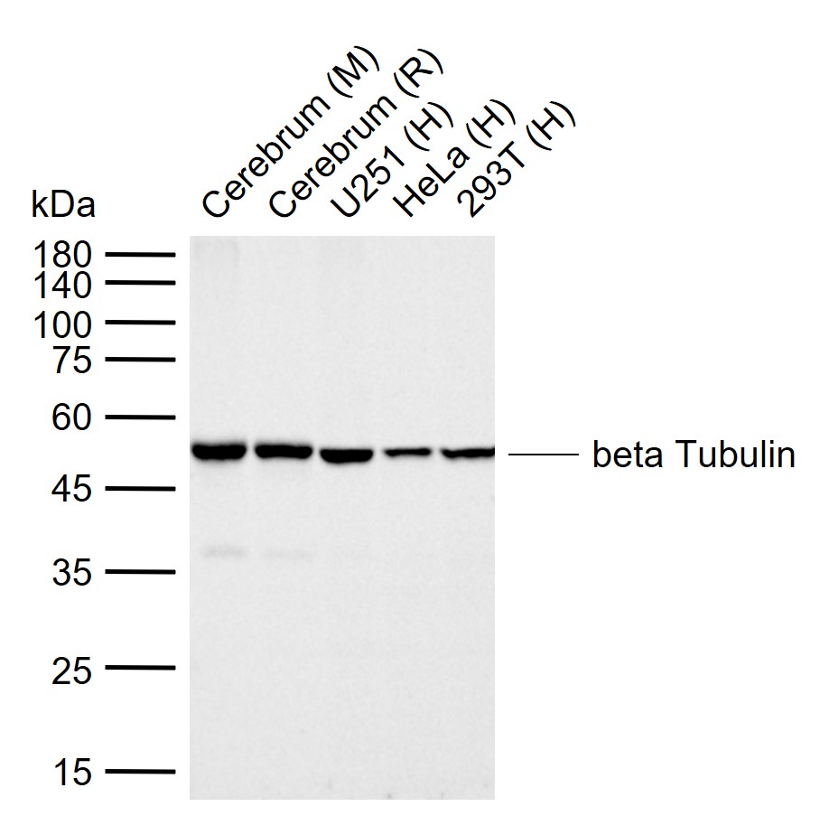
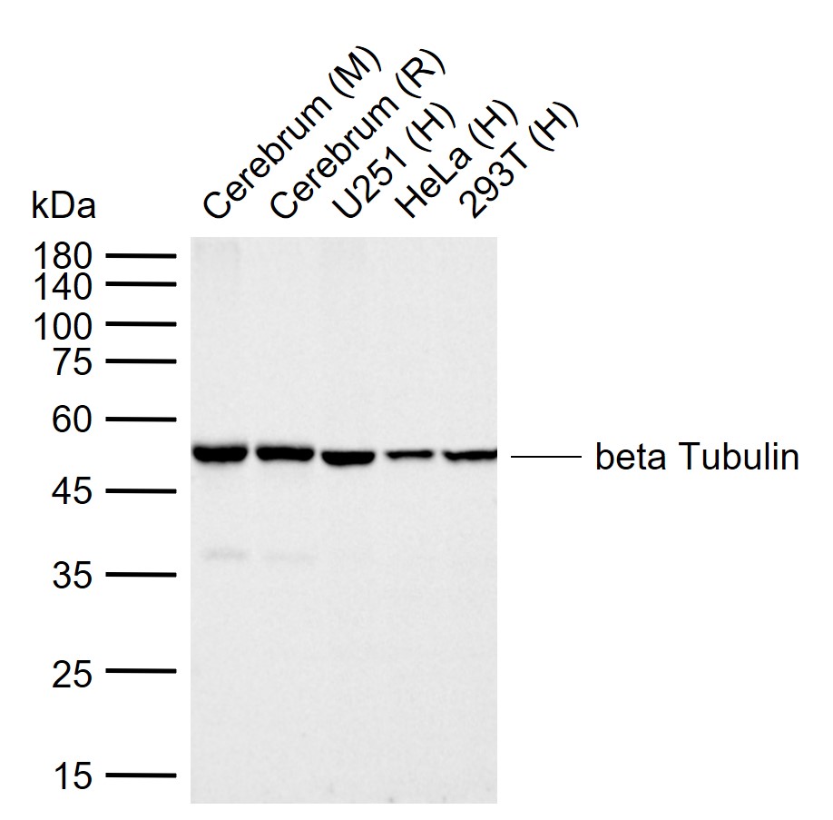
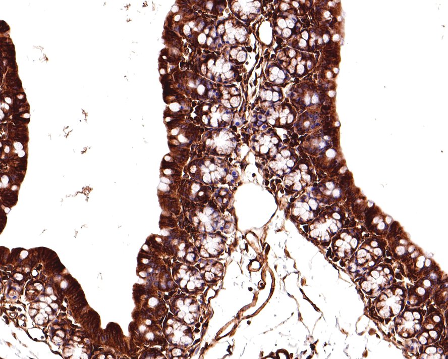
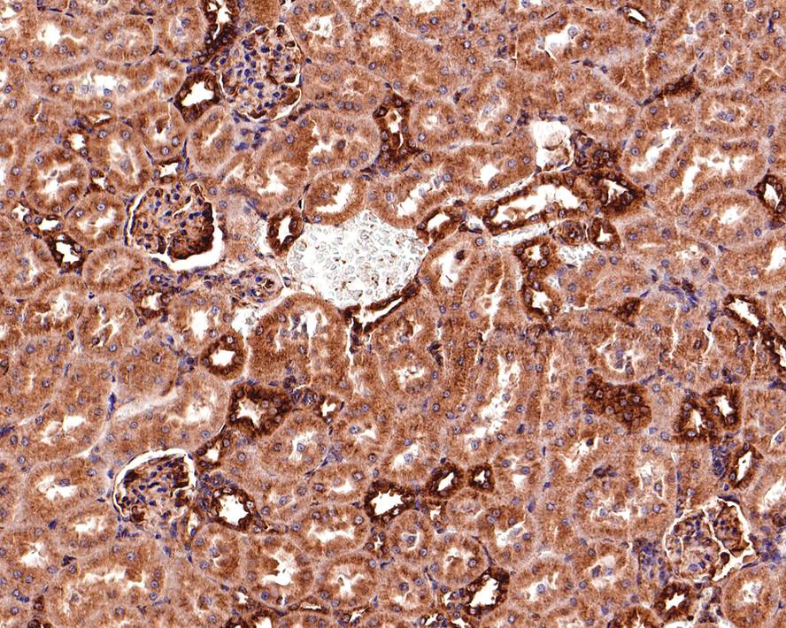
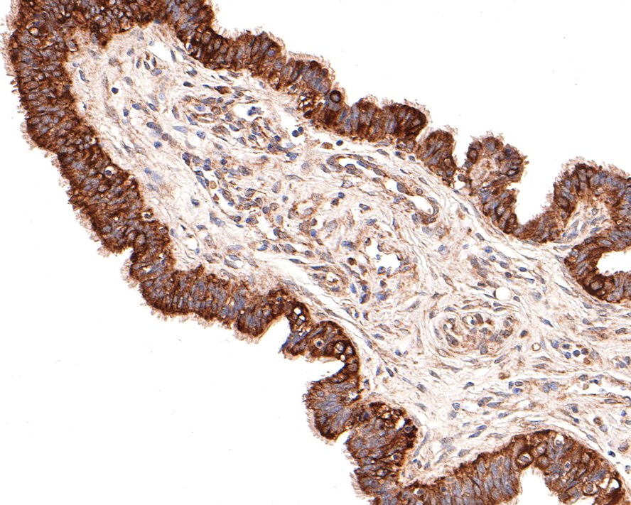
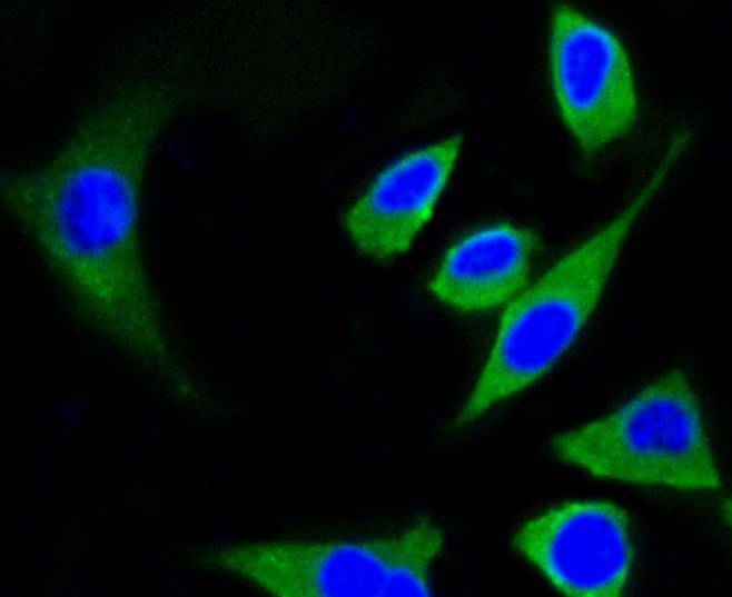
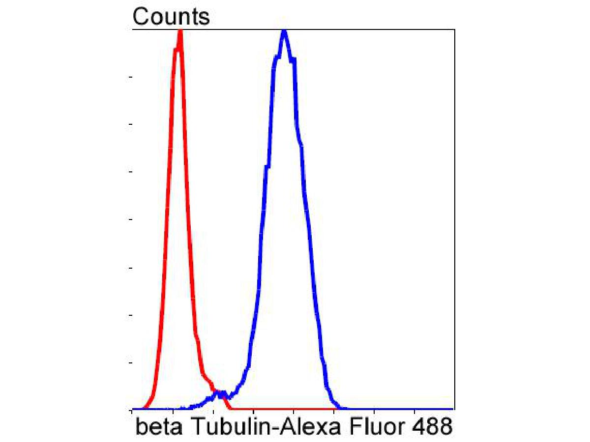
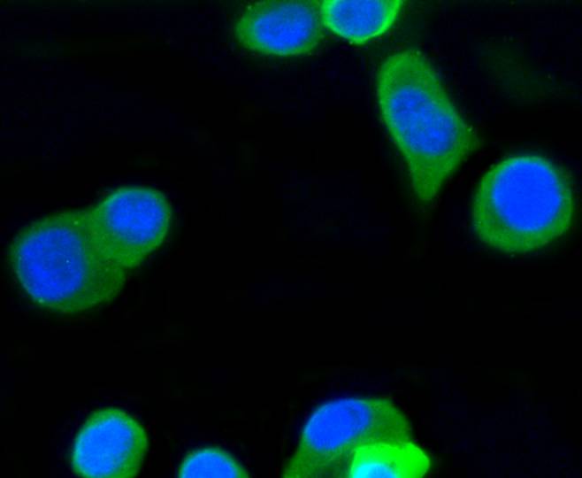
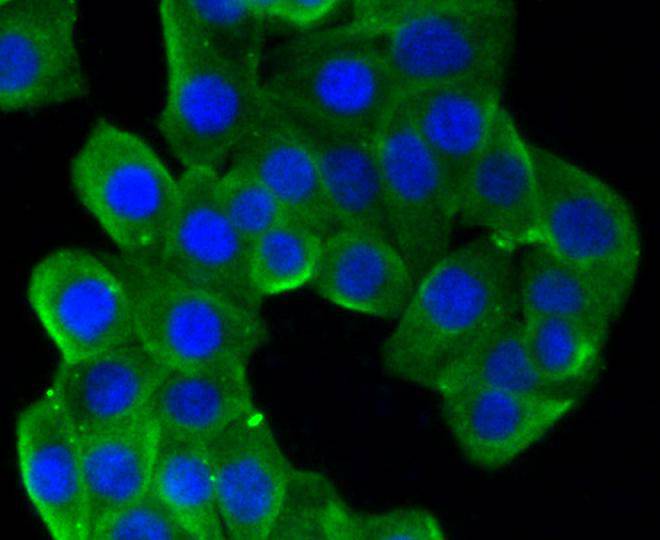
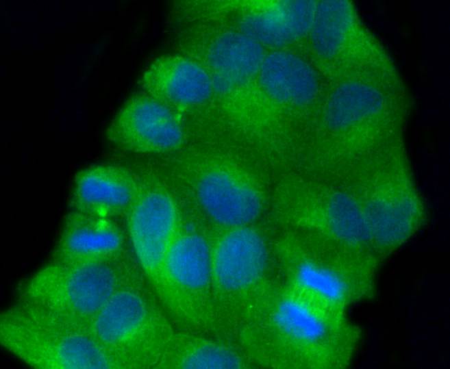
| 產品圖片 | Sample: Lane 1: Mouse Cerebrum tissue lysates Lane 2: Rat Cerebrum tissue lysates Lane 3: Human U251 cell lysates Lane 4: Human HeLa cell lysates Lane 5: Human 293T cell lysates Primary: Anti-beta Tubulin (bsm-52290R) at 1/20000 dilution Secondary: IRDye800CW Goat Anti-Rabbit IgG at 1/20000 dilution Predicted band size: 50 kDa Observed band size: 50 kDa Immunohistochemical analysis of paraffin-embedded mouse large intestine tissue with Rabbit anti-beta Tubulin antibody (bsm-52290R) at 1/400 dilution.The section was pre-treated using heat mediated antigen retrieval with Tris-EDTA buffer (pH 9.0) for 20 minutes. The tissues were blocked in 1% BSA for 20 minutes at room temperature, washed with ddH2O and PBS, and then probed with the primary antibody (bsm-52290R) at 1/400 dilution for 1 hour at room temperature. The detection was performed using an HRP conjugated compact polymer system. DAB was used as the chromogen. Tissues were counterstained with hematoxylin and mounted with DPX. Immunohistochemical analysis of paraffin-embedded rat kidney tissue with Rabbit anti-beta Tubulin antibody (bsm-52290R) at 1/400 dilution.The section was pre-treated using heat mediated antigen retrieval with Tris-EDTA buffer (pH 9.0) for 20 minutes. The tissues were blocked in 1% BSA for 20 minutes at room temperature, washed with ddH2O and PBS, and then probed with the primary antibody (bsm-52290R) at 1/400 dilution for 1 hour at room temperature. The detection was performed using an HRP conjugated compact polymer system. DAB was used as the chromogen. Tissues were counterstained with hematoxylin and mounted with DPX. Immunohistochemical analysis of paraffin-embedded human colon carcinoma tissue with Rabbit anti-beta Tubulin antibody (bsm-52290R) at 1/400 dilution.The section was pre-treated using heat mediated antigen retrieval with Tris-EDTA buffer (pH 9.0) for 20 minutes. The tissues were blocked in 1% BSA for 20 minutes at room temperature, washed with ddH2O and PBS, and then probed with the primary antibody (bsm-52290R) at 1/400 dilution for 1 hour at room temperature. The detection was performed using an HRP conjugated compact polymer system. DAB was used as the chromogen. Tissues were counterstained with hematoxylin and mounted with DPX. Immunohistochemical analysis of paraffin-embedded human fallopian tube tissue with Rabbit anti-beta Tubulin antibody (bsm-52290R) at 1/100 dilution.The section was pre-treated using heat mediated antigen retrieval with Tris-EDTA buffer (pH 9.0) for 20 minutes. The tissues were blocked in 1% BSA for 20 minutes at room temperature, washed with ddH2O and PBS, and then probed with the primary antibody (bsm-52290R) at 1/100 dilution for 1 hour at room temperature. The detection was performed using an HRP conjugated compact polymer system. DAB was used as the chromogen. Tissues were counterstained with hematoxylin and mounted with DPX. ICC staining of beta Tubulin in SH-SY5Y cells (green). Formalin fixed cells were permeabilized with 0.1% Triton X-100 in TBS for 10 minutes at room temperature and blocked with 10% negative goat serum for 15 minutes at room temperature. Cells were probed with the primary antibody (bsm-52290R, 1/50) for 1 hour at room temperature, washed with PBS. Alexa Fluor?488 conjugate-Goat anti-Rabbit IgG was used as the secondary antibody at 1/1,000 dilution. The nuclear counter stain is DAPI (blue). ICC staining of beta Tubulin in PC-12 cells (green). Formalin fixed cells were permeabilized with 0.1% Triton X-100 in TBS for 10 minutes at room temperature and blocked with 10% negative goat serum for 15 minutes at room temperature. Cells were probed with the primary antibody (bsm-52290R, 1/50) for 1 hour at room temperature, washed with PBS. Alexa Fluor?488 conjugate-Goat anti-Rabbit IgG was used as the secondary antibody at 1/1,000 dilution. The nuclear counter stain is DAPI (blue). ICC staining of beta Tubulin in N2A cells (green). Formalin fixed cells were permeabilized with 0.1% Triton X-100 in TBS for 10 minutes at room temperature and blocked with 10% negative goat serum for 15 minutes at room temperature. Cells were probed with the primary antibody (bsm-52290R, 1/50) for 1 hour at room temperature, washed with PBS. Alexa Fluor?488 conjugate-Goat anti-Rabbit IgG was used as the secondary antibody at 1/1,000 dilution. The nuclear counter stain is DAPI (blue). ICC staining of beta Tubulin in SH-SY5Y cells (green). Formalin fixed cells were permeabilized with 0.1% Triton X-100 in TBS for 10 minutes at room temperature and blocked with 10% negative goat serum for 15 minutes at room temperature. Cells were probed with the primary antibody (bsm-52290R, 1/50) for 1 hour at room temperature, washed with PBS. Alexa Fluor?488 conjugate-Goat anti-Rabbit IgG was used as the secondary antibody at 1/1,000 dilution. The nuclear counter stain is DAPI (blue). ICC staining of beta Tubulin in Hela cells (green). Formalin fixed cells were permeabilized with 0.1% Triton X-100 in TBS for 10 minutes at room temperature and blocked with 10% negative goat serum for 15 minutes at room temperature. Cells were probed with the primary antibody (bsm-52290R, 1/50) for 1 hour at room temperature, washed with PBS. Alexa Fluor?488 conjugate-Goat anti-Rabbit IgG was used as the secondary antibody at 1/1,000 dilution. The nuclear counter stain is DAPI (blue). |
我要詢價
*聯系方式:
(可以是QQ、MSN、電子郵箱、電話等,您的聯系方式不會被公開)
*內容:


