| 產(chǎn)品編號 | bs-0029R |
| 英文名稱 | Rabbit Anti-PP2A alpha + beta antibody |
| 中文名稱 | 蛋白質(zhì)磷酸酶-2A抗體 |
| 別 名 | PP2A; PP2A alpha; PP-2A; PP2A C; PP2Ac; PP2CA; PPP2CA; PP2Calpha; RP-C; Protein phosphatase 2, catalytic subunit, alpha isoform; Replication protein C; RP C; PP2AA_HUMAN. PP2A-Cα/β; PP2A-C α/β; PP2A-C α + β; PP2A-C α+β. |
 | Specific References (3) | bs-0029R has been referenced in 3 publications. [IF=3.7] Lin, Lai-xiang, et al. "Feasibility of β-Sheet Breaker Peptide-H102 Treatment for Alzheimers Disease Based on β-Amyloid Hypothesis." PLoS one 9.11 (2014): e112052. IHC-P ; Mouse. [IF=3.33] Zhao, Hai-hua, et al. "Involvement of GSK3 and PP2A in ginsenoside Rb1's attenuation of aluminum-induced tau hyperphosphorylation." Behavioural Brain Research (2012). WB, IHC-P ; Mouse. [IF=1.664] Zhang PF et al. MicroRNA-139 suppresses hepatocellular carcinoma cell proliferation and migration by directly targeting Topoisomerase I. ONCOLOGY LETTERS 17: 1903-1913, 2019 WB ; Human. |
| 研究領(lǐng)域 | 細胞生物 免疫學 信號轉(zhuǎn)導 細胞周期蛋白 激酶和磷酸酶 |
| 抗體來源 | Rabbit |
| 克隆類型 | Polyclonal |
| 交叉反應(yīng) | Human,Mouse,Rat (predicted: Chicken,Dog,Pig,Cow,Rabbit) |
| 產(chǎn)品應(yīng)用 | WB=1:500-2000, IHC-P=1:100-500, IHC-F=1:100-500, IF=1:100-500, Flow-Cyt=1ug/Test, ELISA=1:5000-10000 not yet tested in other applications. optimal dilutions/concentrations should be determined by the end user. |
| 理論分子量 | 34kDa |
| 細胞定位 | 細胞核 細胞漿 |
| 性 狀 | Liquid |
| 濃 度 | 1mg/ml |
| 免 疫 原 | KLH conjugated synthetic peptide derived from human PP-2A: 205-309/309 |
| 亞 型 | IgG |
| 純化方法 | affinity purified by Protein A |
| 緩 沖 液 | 0.01M TBS(pH7.4) with 1% BSA, 0.03% Proclin300 and 50% Glycerol. |
| 保存條件 | Shipped at 4℃. Store at -20 °C for one year. Avoid repeated freeze/thaw cycles. |
| 注意事項 | This product as supplied is intended for research use only, not for use in human, therapeutic or diagnostic applications. |
| PubMed | PubMed |
| 產(chǎn)品介紹 | This gene encodes the phosphatase 2A catalytic subunit. Protein phosphatase 2A is one of the four major Ser/Thr phosphatases, and it is implicated in the negative control of cell growth and division. It consists of a common heteromeric core enzyme, which is composed of a catalytic subunit and a constant regulatory subunit, that associates with a variety of regulatory subunits. This gene encodes an alpha isoform of the catalytic subunit. [provided by RefSeq, Jul 2008]. This antibody is crosed with PP-2A subunit A,B. Function: PP2A can modulate the activity of phosphorylase B kinase casein kinase 2, mitogen-stimulated S6 kinase, and MAP-2 kinase. Cooperates with SGOL2 to protect centromeric cohesin from separase-mediated cleavage in oocytes specifically during meiosis I (By similarity). Can dephosphorylate SV40 large T antigen and p53/TP53. Dephosphorylates SV40 large T antigen, preferentially on serine residues 120, 123, 677, and perhaps 679. The C subunit was most active, followed by the AC form, which was more active than the ABC form, and activity of all three forms was strongly stimulated by manganese, and to a lesser extent by magnesium. Dephosphorylation by the AC form, but not C or ABC form is inhibited by small T antigen. Activates RAF1 by dephosphorylating it at 'Ser-259'. Subunit: PP2A consists of a common heterodimeric core enzyme, composed of PPP2CA a 36 kDa catalytic subunit (subunit C) and PPP2R1A a 65 kDa constant regulatory subunit (PR65 or subunit A), that associates with a variety of regulatory subunits. Proteins that associate with the core dimer include three families of regulatory subunits B (the R2/B/PR55/B55, R3/B''/PR72/PR130/PR59 and R5/B'/B56 families), the 48 kDa variable regulatory subunit, viral proteins, and cell signaling molecules. Interacts with NXN; the interaction is direct (By similarity). Interacts with TP53, SGOL1 and SGOL2. Interacts with AXIN1; the interaction dephosphorylates AXIN1. Interacts with PIM3; this interaction promotes dephosphorylation, ubiquitination and proteasomal degradation of PIM3. Interacts with RAF1. Subcellular Location: Cytoplasm. Nucleus. Chromosome, centromere. Cytoplasm, cytoskeleton, spindle pole. Note=In prometaphase cells, but not in anaphase cells, localizes at centromeres. During mitosis, also found at spindle poles. Centromeric localization requires the presence of SGOL2 (By similarity). Post-translational modifications: Reversibly methyl esterified on Leu-309. Carboxyl methylation may play a role in holoenzyme assembly, enhancing the affinity of the PP2A core enzyme for some, but not all, regulatory subunits. It varies during the cell cycle. Phosphorylation of either threonine (by autophosphorylation-activated protein kinase) or tyrosine results in inactivation of the phosphatase. Auto-dephosphorylation has been suggested as a mechanism for reactivation. Similarity: Belongs to the PPP phosphatase family. PP-1 subfamily. SWISS: P67775 Gene ID: 5515 Database links: Entrez Gene: 416318 Chicken Entrez Gene: 5515 Human Entrez Gene: 19052 Mouse Entrez Gene: 100009252 Rabbit Omim: 176915 Human SwissProt: P48463 Chicken SwissProt: P67775 Human SwissProt: P63330 Mouse SwissProt: P67777 Rabbit Unigene: 105818 Human Unigene: 260288 Mouse Unigene: 1271 Rat 激酶和磷酸酶(Kinases and Phosphatases) PP-2A (protein phosphatase 2A catalytic subunit;PP2A alpha;)參與酵母細胞及兩棲類卵母細胞有絲分裂的蛋白絲/蘇氨酸磷酸酶。 此酶的表達與細胞周期調(diào)節(jié)有關(guān)。此抗體與PP-2A subunit A,B,C 均有交叉反應(yīng)。 |
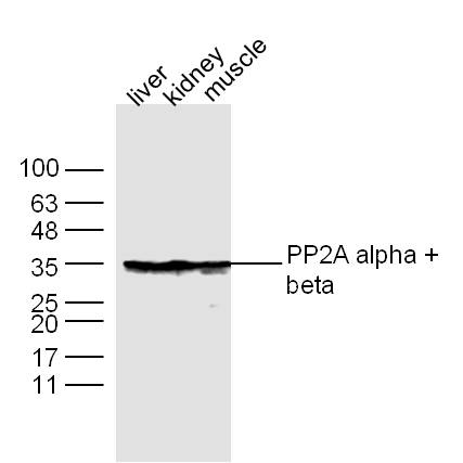
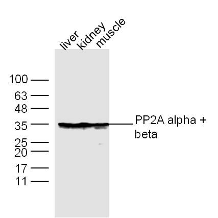
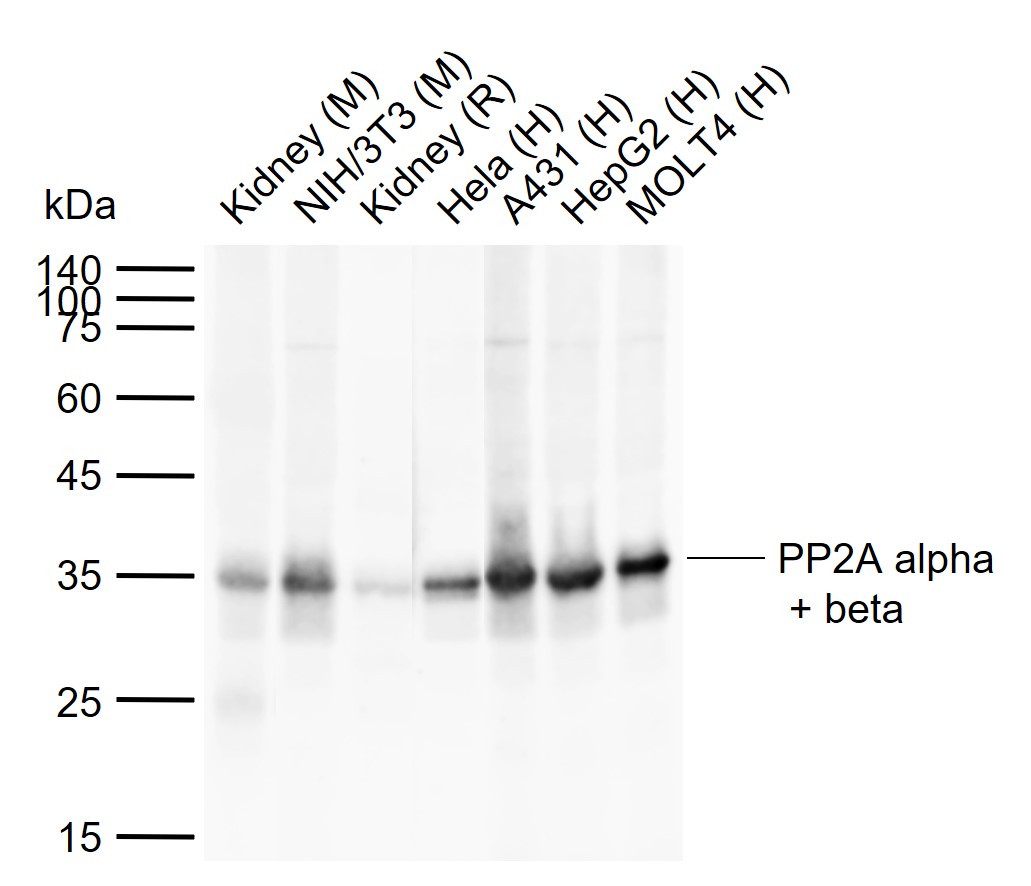
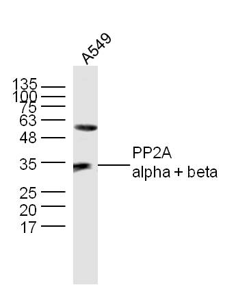
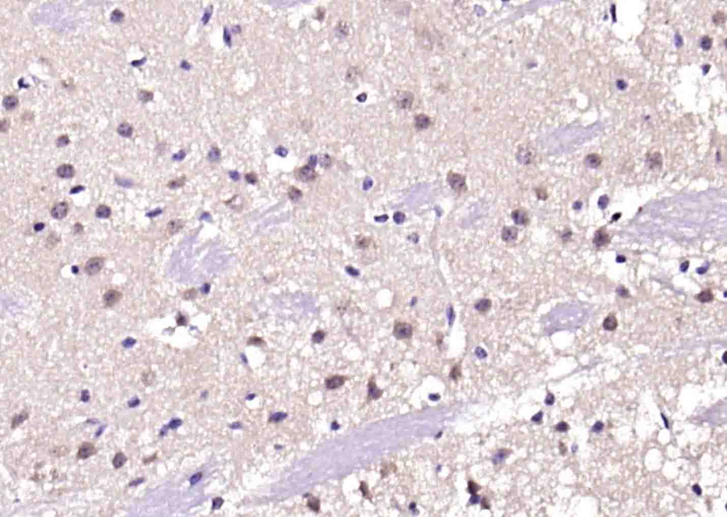
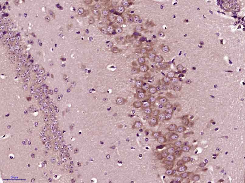
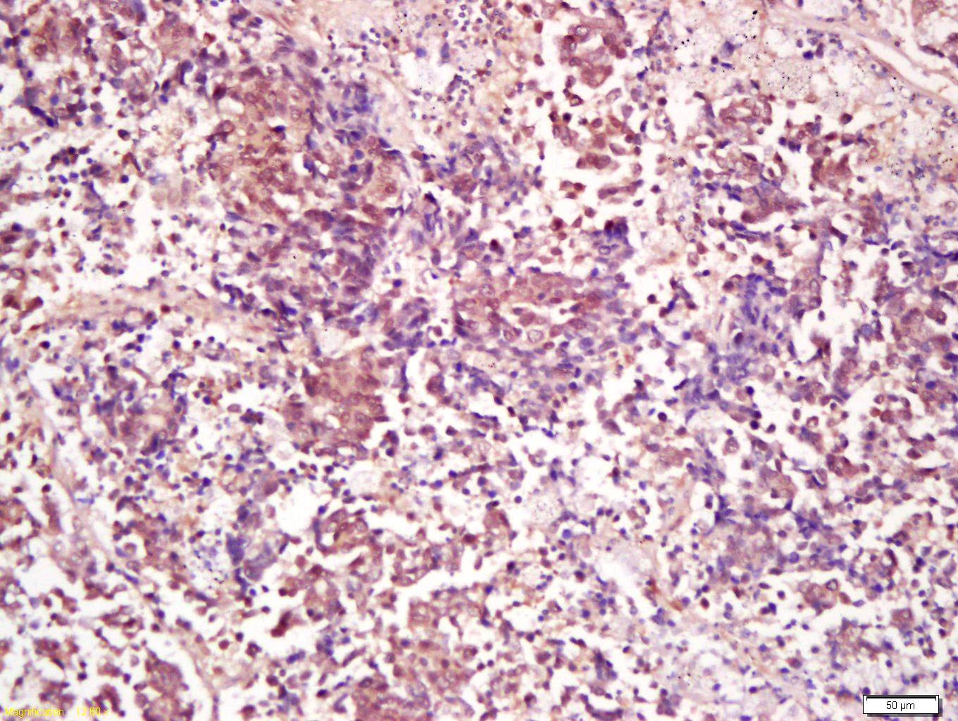
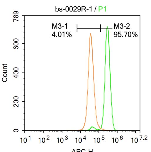
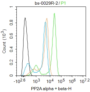
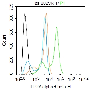
| 產(chǎn)品圖片 | Sample: Liver (Mouse) Lysate at 30 ug Kidney (Mouse) Lysate at 30 ug Muscle (Mouse) Lysate at 30 ug Primary: Anti- PP2A alpha + beta (bs-0029R) at 1/300 dilution Secondary: IRDye800CW Goat Anti-Rabbit IgG at 1/20000 dilution Predicted band size: 34 kD Observed band size: 34 kD Sample: Lane 1: Mouse Kidney tissue lysates Lane 2: Mouse NIH/3T3 cell lysates Lane 3: Rat Kidney tissue lysates Lane 4: Human Hela cell lysates Lane 5: Human A431 cell lysates Lane 6: Human HepG2 cell lysates Lane 7: Human MOLT4 cell lysates Primary: Anti-PP2A alpha + beta (bs-0029R) at 1/1000 dilution Secondary: IRDye800CW Goat Anti-Rabbit IgG at 1/20000 dilution Predicted band size: 34 kDa Observed band size: 34 kDa Sample: A549 Cell (Human) Lysate at 30 ug Primary: Anti-PP2A alpha + beta (Bs- 0029R) at 1/300 dilution Secondary: IRDye800CW Goat Anti-Rabbit IgG at 1/20000 dilution Predicted band size: 34 kD Observed band size: 34 kD Paraformaldehyde-fixed, paraffin embedded (mouse brain); Antigen retrieval by boiling in sodium citrate buffer (pH6.0) for 15min; Block endogenous peroxidase by 3% hydrogen peroxide for 20 minutes; Blocking buffer (normal goat serum) at 37°C for 30min; Antibody incubation with (PP2A alpha + beta) Polyclonal Antibody, Unconjugated (bs-0029R) at 1:200 overnight at 4°C, followed by operating according to SP Kit(Rabbit) (sp-0023) instructionsand DAB staining. Paraformaldehyde-fixed, paraffin embedded (Mouse brain); Antigen retrieval by boiling in sodium citrate buffer (pH6.0) for 15min; Block endogenous peroxidase by 3% hydrogen peroxide for 20 minutes; Blocking buffer (normal goat serum) at 37°C for 30min; Antibody incubation with (PP2A alpha + beta) Polyclonal Antibody, Unconjugated (bs-0029R) at 1:400 overnight at 4°C, followed by operating according to SP Kit(Rabbit) (sp-0023) instructionsand DAB staining. Tissue/cell: human lung carcinoma; 4% Paraformaldehyde-fixed and paraffin-embedded; Antigen retrieval: citrate buffer ( 0.01M, pH 6.0 ), Boiling bathing for 15min; Block endogenous peroxidase by 3% Hydrogen peroxide for 30min; Blocking buffer (normal goat serum,C-0005) at 37℃ for 20 min; Incubation: Anti-PP2A alpha+beta Polyclonal Antibody, Unconjugated(bs-0029R) 1:200, overnight at 4°C, followed by conjugation to the secondary antibody(SP-0023) and DAB(C-0010) staining Blank control: A431. Primary Antibody (green line): Rabbit Anti-PP2A alpha + beta antibody (bs-0029R) Dilution: 1μg /10^6 cells; Isotype Control Antibody (orange line): Rabbit IgG . Secondary Antibody : Goat anti-rabbit IgG-AF647 Dilution: 1μg /test. Protocol The cells were fixed with 4% PFA (10min at room temperature)and then permeabilized with 90% ice-cold methanol for 20 min at-20℃. The cells were then incubated in 5%BSA to block non-specific protein-protein interactions for 30 min at at room temperature .Cells stained with Primary Antibody for 30 min at room temperature. The secondary antibody used for 40 min at room temperature. Acquisition of 20,000 events was performed. Blank control: Hela. Primary Antibody (green line): Rabbit Anti-PP2A alpha + beta antibody (bs-0029R) Dilution: 2ug/Test; Secondary Antibody : Goat anti-rabbit IgG-FITC Dilution: 0.5ug/Test. Protocol The cells were fixed with 4% PFA (10min at room temperature)and then permeabilized with 90% ice-cold methanol for 20 min at -20℃.The cells were then incubated in 5%BSA to block non-specific protein-protein interactions for 30 min at room temperature .Cells stained with Primary Antibody for 30 min at room temperature. The secondary antibody used for 40 min at room temperature. Acquisition of 20,000 events was performed. Blank control: Hela. Primary Antibody (green line): Rabbit Anti-PP2A alpha + beta antibody (bs-0029R) Dilution: 1ug/Test; Secondary Antibody : Goat anti-rabbit IgG-FITC Dilution: 0.5ug/Test. Protocol The cells were fixed with 4% PFA (10min at room temperature)and then permeabilized with 90% ice-cold methanol for 20 min at -20℃.The cells were then incubated in 5%BSA to block non-specific protein-protein interactions for 30 min at room temperature .Cells stained with Primary Antibody for 30 min at room temperature. The secondary antibody used for 40 min at room temperature. Acquisition of 20,000 events was performed. |
我要詢價
*聯(lián)系方式:
(可以是QQ、MSN、電子郵箱、電話等,您的聯(lián)系方式不會被公開)
*內(nèi)容:


